
More Information
Submitted: May 25, 2026 | Accepted: June 04, 2026 | Published: June 05, 2026
Citation: lam M, Khan AA, Shabnam MS, Hasan SM, Anas M, Zaidi SMH. Development of Thiadiazole-Based VEGFR-2 Targeted Agents: A Comprehensive Review of Cytotoxic and Anticancer Activities. Arch Pharm Pharma Sci. 2026; 10(1): 5-20. Available from:
https://dx.doi.org/10.29328/journal.apps.1001071
DOI: 10.29328/journal.apps.1001071
Copyright license: © 2026 Alam M, et al, et al. This is an open access article distributed under the Creative Commons Attribution License, which permits unrestricted use, distribution, and reproduction inany medium, provided the original work is properly cited.
Keywords: 1,3,4-Thiadiazole derivatives; VEGFR-2 inhibitors; Anticancer agents; Tumor angiogenesis; Structure-activity relationships; Cytotoxicity evaluation; Apoptosis induction

Development of Thiadiazole-Based VEGFR-2 Targeted Agents: A Comprehensive Review of Cytotoxic and Anticancer Activities
Mehtab Alam1, Ahsan Ahmad Khan1*, Mir Snober Shabnam2, Syed Misbahul Hasan1, Mohd Anas1 and Syed Mehndi Hasan Zaidi1
1Department of Pharmaceutical Chemistry, Faculty of Pharmacy, Integral University, Kursi Road, Lucknow-226026, Uttar Pradesh, India
2Department of Bioscience, Integral University, Kursi Road, Lucknow-226026, Uttar Pradesh, India
*Corresponding author: Dr. Ahsan Ahmad Khan, Associate Professor, Faculty of Pharmacy, Integral University, Kursi Road, Lucknow, Uttar Pradesh, 226026, India, Email: [email protected]
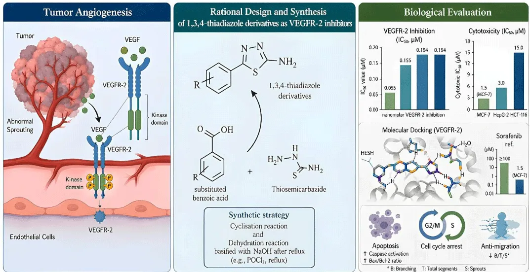
One of the best-proven molecular targets in the treatment of cancer is vascular endothelial growth factor receptor-2 (VEGFR-2). Because it plays an important role in tumor angiogenesis, proliferation, and metastasis. FDA-approved VEGFR-2 inhibitors have proven to have clinical efficacy in managing various malignancies. Anti-cancer activity of approved agents such as sorafenib (IC50 = 1 µM - 10 µM), sunitinib (IC50 = 1 µM - 5 µM), and pazopanib (IC50 = 1 µM - 3 µM) has been demonstrated. However, their therapeutic use can result in off-target toxicities. Moreover, acquired resistance mechanisms have been observed as well. They also have poorly selective means of action. The 1,3,4-thiadiazole scaffold has become an important heterocyclic compound of medicinal chemistry due to its mesoionic character, versatility in structures, and pharmacological attributes. This review presents a systematic study of the earlier developments in the rational design, synthesis, SAR, and cytotoxicity evaluation of Thiadiazole-based VEGFR-2 inhibitors. In this review, the molecular mechanisms of VEGFR-2 inhibitors and their biological activities against a variety of cancer cell lines in terms of their ability to induce apoptosis, modulate cell cycle, anti-metastatic properties, and in silico approaches. The recently added Thiadiazole derivatives display very good VEGFR-2 inhibitory activities with IC50 values ranging from 0.055 to 0.194 µM, potent antiproliferation activities with IC50 values of 1.5-15 µM, good selectivity indices (3-20-fold), and anticancer activities through different mechanisms of action that are more favorable than the established drugs.
Graphical Abstract
Cancer is one of the most urgent social health emergencies in the whole world and especially in India, as the World Health Organization (WHO) GLOBOCAN 2025 report offers the central estimates according to the 2022 updates and forecasts. In the world, it was estimated in the year 2022 that there were about 20 million new cases of cancer resulting in about 9.7 million deaths, and it is projected that this figure will increase drastically to 35 million new cases by the year 2050. This epidemic is due to the aging of the world population, increased incidence of preventable risk factors such as smoking of tobacco, alcohol, poor nutrition, sedentary lifestyles, obesity, and environmental pollutants such as air pollution, and the chronic infectious agents like human papillomavirus (HPV), hepatitis B and C viruses, and Helicobacter pylori. Cancer related to lifestyle is predominant in high-income nations; infection contributes to it in much greater proportions in low- and middle-income nations, which incur 70 per cent of the world’s cancer burden. WHO reported the different types of cancers spread worldwide with their numbers by 2025, as shown in Figure 1 [1,2].
Figure 1: Percentage of cancer by WHO 2025.
In India, the disease has also become critically worrying as the number of new cases and deaths in 2022 is estimated at 1.41 million and over 1 million, respectively, placing it as the third-highest absolute incidence and the second-highest mortality despite its status as home to only 18% of the world population, but with 8.8% of cases. This unequal distribution is due to the combination of local risk factors such as high levels of tobacco consumption (both smoked (bidis) and smokeless (gutkha) which is linked to 30-50% of male cancers) and prevalence of HPV/hepatitis infections with low levels of vaccination coverage, alcohol use, nutritional deficiencies, poverty-oriented late-stage manifestations, and insufficient screening infrastructure which have led to a five-year survival rate of less than 30% compared to over 60 years in high-income countries The cancer profile in India does not follow global trends, and reflects the differences in socio cultural and epidemiological terms: breast cancer is the leading at 13.6% (rapidly increasing with urbanization, change in diet to processed food and consequence with delayed child bearing) and lip and oral cavity at 10.2% (India has contributed nearly half of the oral cancer cases in the world, with most of them due to betel quid chewing in combination with tobacco), cervix uteri cancer at 9.0 and in esophagus cancer at 5.0 The regional differences are stark, oral cancers in the northeast through habits of eating the areca nut, cervical in the south through lower awareness, and breast/lung in the urban areas, and compounded by over stretched health care systems and rural-urban differences (90% of the population, minimal services in the rural) versus urban (better access) [3,4].
Expanded global trends
It has been projected that there will be an increase of 77% in the number of low-human development index (HDI) countries by 2050, and hence there is a need to ensure equitable access to preventive, early-detection, radiotherapy (a 77% deficiency in low-HDI areas), and affordable treatments.
India challenges and variations
According to the National Cancer Registry Programme (NCRP) data on 43 registries, tobacco predominates in men (45% attribution) and infections in women (25%), and the Ayushman Bharat program is trying to close the gap but is experiencing challenges in implementation (Figure 2).
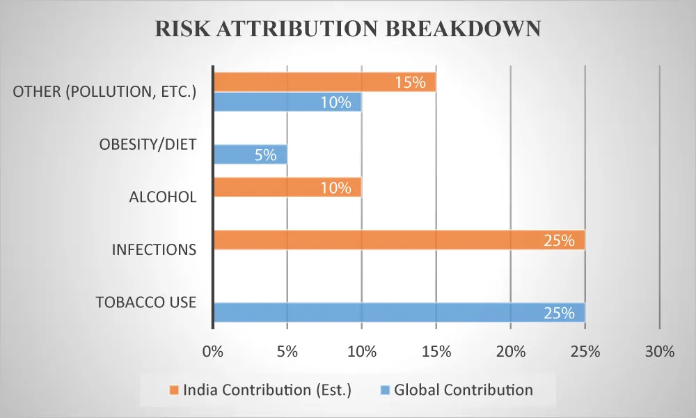
Figure 2: Risk Attribution Breakdown.
Angiogenesis
Angiogenesis refers to the process of formation of new blood vessels from existing ones, as shown in Figure 3. It is an essential biological process that gets critically deregulated in cancer. According to Hanahan and Weinberg, various cancer hallmarks, sustained angiogenesis is one such hallmark which leads to the supply of vascular oxygen for the rapid growth of the tumor and the spread of metastasis. The vascular system of a tumor allows nutrient, oxygen, and immune cell transfer and is regulated by pro- and anti-angiogenic factors working in balance [5,6].
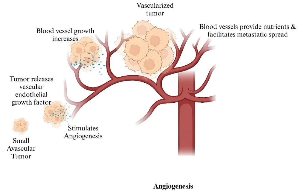
Figure 3: Tumor angiogenesis.
Around 40 years ago, the discovery of the vascular permeability and angiogenesis-promoting factor led to the identification of VEGF family ligands and receptors (VEGFRs). The VEGF/VEGFR axis is essential for angiogenesis and is recognized as a primary mediator of tumor blood vessel development [7,8].
The tumor angiogenic pathways function through diverse mechanisms, including sprouting angiogenesis, intussusceptive angiogenesis, vascular co-option, vascular mimicry, and glomeruloid angiogenesis, which are normally activated by varied angiogenic stimulators and their receptors [9,10]. Among the three main VEGF receptors, VEGFR-1, VEGFR-2, and VEGFR-3, as shown in the given (Figure 4), VEGFR-2 emerges as one of the key and critical mediators in tumor angiogenesis and is recognized as a major therapeutic target for combating the angiogenesis phenomenon [11,12].
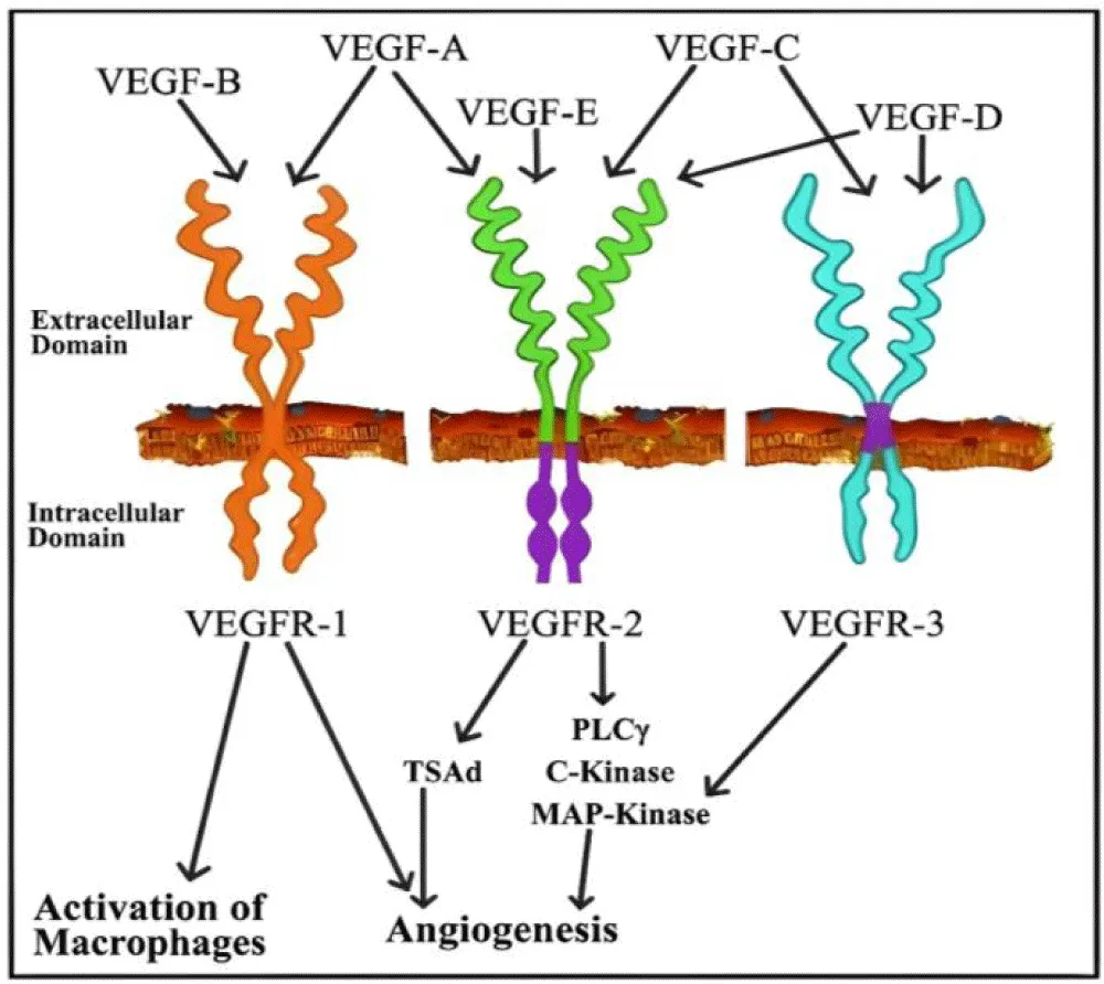
Figure 4: Types of VEGFR receptors.
The growth, survival, and metastasis of solid tumors depend on angiogenesis. Moreover, VEGFR-2, which is the receptor for VEGF, is overexpressed in many human solid tumors. Thus, it makes VEGFR-2 an attractive target for anti-cancer therapy. The protein VEGFR-2 is involved in tumor angiogenesis, and its high expression in a variety of tumor types has made it a target for anticancer therapy [13,14]. VEGFs are vascular endothelial growth factors that are responsible for directing angiogenesis and vasculogenesis. VEGFR-2 is the receptor of VEGF and during various physiological reaction it regulate response of the receptor. VEGF is mainly expressed in endothelial cells and performs cell differentiation, cell proliferation, migration, and survival [15].
VEGFR-2: Molecular architecture and clinical validation
VEGFR-2 is a receptor tyrosine kinase that mediates the effect of VEGF in angiogenesis, as the major receptor of VEGF. It is also called KDR (Kinase insert Domain Receptor) or Flk1. Receptor tyrosine kinases are widely conserved molecules ranging from a prokaryote to man, which take an active part in the phosphorylation of the tyrosine residues in the proteins, resulting in alterations in protein function. Deregulation of kinases due to mutation and transcriptional or post-translational modifications ultimately leads to the onset of pathological conditions, including cancer [16,17]. Among the various kinases, the VEGF/VEGFR-2 signaling cascade is an important target to develop novel small-molecule inhibitors for the therapy of abnormal angiogenesis associated with cancer. Due to advances in the knowledge of the catalytic domain and DFG-motif (Aspartate-Phenylalanine-Glycine) region, selective DFG-in (type I) and DFG-out (type II) VEGFR-2/KDR inhibitors were successfully developed, and some are in different phases of clinical trials. The DFG-out (inactive) conformation has significant advantages over the DFG-in (active) conformation concerning the affinity of ATP at the catalytic domain [18,19].
The clinical validation of VEGFR-2 as a therapeutic target is evidenced by multiple FDA-approved inhibitors. Several anti-angiogenic drugs, like ramucirumab, sunitinib, axitinib, and sorafenib, showing good survival rates, have been developed and FDA-approved against VEGFR-2 [20,21].However, clinical application of available VEGFR-2 inhibitors has been challenged by limited efficacy and a wide range of side effects, potentially due to inadequate selectivity for VEGFR-2. Analysis of residual kinase activity of a panel of 270 kinases showed that highly selective VEGFR-2 inhibitors displayed greater selectivity compared with reference inhibitors, highlighting that toxicities associated with available VEGFR-2 inhibitors are thought to be partly due to their effects against kinases other than VEGFR-2 [22].
The clinical utility of VEGFR-2 inhibition has been firmly established through decades of controlled trials, with approved agents now spanning multiple tumor types and lines of therapy. Among the small-molecule inhibitors, sorafenib holds particular historical significance as one of the earliest multi-kinase agents to demonstrate survival benefit in solid tumors, earning regulatory approval for unresectable hepatocellular carcinoma, advanced renal cell carcinoma, and radioiodine-refractory differentiated thyroid carcinoma [23]. Sunitinib, similarly broad in its kinase coverage, extended this paradigm by showing meaningful progression-free survival gains in advanced renal cell carcinoma and subsequently in imatinib-resistant gastrointestinal stromal tumors and progressive pancreatic neuroendocrine tumors, establishing multi-targeted VEGFR inhibition as a viable first-line strategy in these settings [24]. Axitinib emerged as a more selective second-generation agent, designed specifically to address the shortcomings of earlier inhibitors; it is now standard of care for advanced renal cell carcinoma following failure of at least one prior systemic regimen, with particular benefit observed in patients whose disease progressed on sunitinib or cytokine-based therapies [25]. Pazopanib, with clinical outcomes comparable to sunitinib in treatment-naive renal cell carcinoma, extended the therapeutic reach of VEGFR-2 blockade into soft tissue sarcomas refractory to prior chemotherapy, demonstrating that anti-angiogenic strategies are not exclusively confined to epithelial malignancies [26]. Lenvatinib, a structurally distinct inhibitor with a broader receptor kinase profile encompassing VEGFR-1/2/3, FGFR1–4, RET, KIT, and PDGFRα, has since been approved across radioiodine-refractory thyroid cancer, unresectable hepatocellular carcinoma, and advanced renal cell carcinoma in combination regimens with everolimus and pembrolizumab, underscoring the expanding role of VEGFR-targeted agents within immuno-oncology combinations. Cabozantinib introduced a mechanistically important refinement by simultaneously targeting VEGFR-2, MET, and AXL — receptor axes that are frequently co-activated during acquired resistance to first-generation anti-angiogenic therapy — and has demonstrated efficacy in advanced renal cell carcinoma, hepatocellular carcinoma, and differentiated thyroid cancer following prior treatment [27]. Beyond small molecules, ramucirumab, a fully human monoclonal antibody directed against the extracellular ligand-binding domain of VEGFR-2, has validated receptor-level blockade as a distinct and effective approach, with approvals spanning gastric and gastro-oesophageal junction adenocarcinoma, metastatic non-small cell lung cancer, colorectal cancer, and hepatocellular carcinoma [28]. Taken together, the patient populations deriving the most consistent benefit from VEGFR-2-directed therapy include those with clear-cell renal cell carcinoma histology, tumors characterized by elevated VEGF/VEGFR-2 pathway activity, and patients whose disease has progressed beyond first-line cytotoxic or targeted regimens — a clinical reality that underscores the urgent need for structurally novel inhibitors capable of improved selectivity and a more favorable tolerability profile. It is precisely against this backdrop that the rational development of thiazole-based VEGFR-2 inhibitors acquires its clinical significance.
The 1,3,4-Thiadiazole Scaffold in Drug Discovery: The impact of heterocycles cannot be understated. Medicinal chemistry contains numerous drug molecules based on heterocyclic structures. In fact, 75% of the drug molecules approved by the FDA and still available on the market contain heterocycles. Over the next ten years, a far larger proportion of novel drugs that will incorporate nitrogen-containing heterocyclic structures is forecasted. Five-membered heterocycles with two heteroatoms, mainly nitrogen and sulphur, have gained a lot of attention for their biological properties and good drug-like properties [29,30].
The 1,3,4-thiadiazole scaffold has emerged as a privileged moiety in cancer drug discovery due to its mesoionic character, structural diversity, and molecular pharmacology. Notably, the N-N-C-S motif and sulfur atom of thiadiazole significantly contribute to VEGFR-2 binding through key molecular interactions. A systematic analysis of different publications from the last decade led to the extraction and evaluation of thiazole-based VEGFR-2 inhibitors, with chemical space, structure-activity relationships as shown below (Figure 5), substitution patterns, selectivity, toxicity, and essential binding interactions (ATP or allosteric site) with VEGFR-2 being critically examined [31,32].

Figure 5: SAR of thiadiazole.
The chemistry of 1,3,4-thiadiazole is one of the most interesting scaffolds for synthesizing new drug molecules due to its numerous pharmacological activities. Several modifications in the thiazole ring have been made, proving it to be more potent and highly effective with a less toxic scaffold for various biological applications [33]. During recent years, small molecules containing five-member heterocyclic moieties have become the subject of considerable growing interest for designing new antitumor agents, with 1,3,4-thiadiazole being one of [34,35].
Rationale, scope, and organization
This comprehensive review addresses the critical need for selective and efficacious VEGFR-2 inhibitors by systematically analyzing recent advances in thiazole-based inhibitor development. The review is structured to give: thorough coverage of VEGFR-2 structural biology, signaling pathways, and role in cancer, in-depth analysis of 1,3,4-thiadiazole chemistry, synthetic strategies and structural diversity, a systematic overview of recent reports of thiadiazole-based VEGFR-2 inhibitors including design strategies, synthetic pathways, and biological testing, and extensive structure-activity relationship investigations [36,37]. Extensive cytotoxicity studies across various cancer cell lines with selectivity studies; mechanistic studies such as apoptosis pathways, cell cycle regulation, and anti-metastatic effects; future outlook and translational prospects [38].
VEGFR-2: Structural biology, signaling mechanisms, and cancer biology molecular architecture and structural features of VEGFR-2
The conserved three-dimensional structure of receptor tyrosine kinases (RTKs) has been observed in prokaryotes to humans and actively participates in the phosphorylation process of tyrosine residues in proteins, which results in the alteration of protein function. The human genome encodes two kinds of tyrosine kinases: non-receptor tyrosine kinases (NRTKs) and receptor tyrosine kinases (RTKs), with VEGFR-2 belonging to the RTK superfamily [39,40].
VEGFR-2 is a receptor that regulates the process of vasculogenesis and angiogenesis through catalytic receptor tyrosine kinases. The structure of the protein has three major structural domains. They include an extracellular ligand-binding domain having seven immunoglobulin (Ig)-like domains (D1-D7), a single-pass transmembrane helix, and an intracellular segment with the tyrosine kinase catalytic domain, as shown in the given figure (Figure 6) [41,42].According to the research, D2 and D3 are the primary binding sites for VEGF-A, and the ligand with a high affinity binds to it via a sandwich-like molecular structure [43-45].
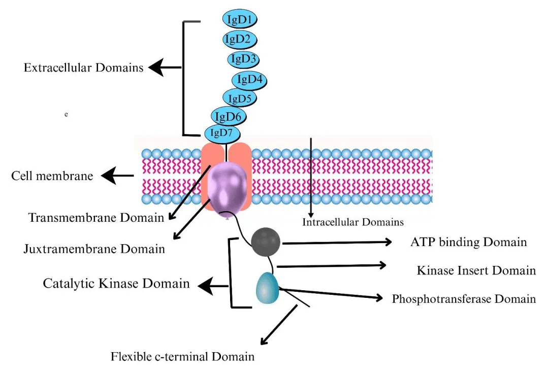
Figure 6: Molecular architecture of VEGFR-2.
The intracellular tyrosine kinase domain contains the ATP-binding site, activation loop, and multiple tyrosine residues that undergo autophosphorylation upon receptor activation. In the catalytic domain, between the front and back cleft, a smaller gatekeeper residue (Val916) is present; therefore, selectivity against VEGFR-2 could be precisely achieved [46,47]. This structural feature distinguishes VEGFR-2 from many other kinases that possess larger gatekeeper residues, creating opportunities for developing selective inhibitors [48,49].
The DFG motif, consisting of Asp-Phe-Gly residues, serves as a molecular switch controlling the kinase activation state. Small molecule first-generation type I, DFG-in, and second-generation type II, DFG-out, VEGFR-2 inhibitors exhibit clinical benefits in the treatment of aberrant angiogenesis associated with cancer [50,51].
VEGF-mediated VEGFR-2 activation and signal transduction
The VEGFs and their VEGFRs (receptors) have critical roles in vasculogenesis and angiogenesis. Angiogenesis is a major mechanism involved in many physiological and pathological processes. It plays an important role in the proliferation, migration, and survival of endothelial cells, which further leads to tubulogenesis and finally the formation of vessels. The VEGF/VEGFR-2 system precisely controls the signaling cascade pathways in this series [52,53].
The VEGF binding to the IgD2 and IgD3 domains of VEGFR-2 induces the dimerization of the receptor, subsequently causing activation and trans-autophosphorylation of the tyrosine kinase, and then initiating intracellular signaling cascades. Finally, the VEGF-activated VEGFR-2 stimulates and mediates a variety of signaling transduction, biological responses, and pathological processes in angiogenesis [54,55].
Several crucial phosphorylated sites within the VEGFR-2 intracellular domains mediate several key signaling processes. Tyr801, Tyr951, Tyr1175, and Tyr1214 in the VEGFR-2 intracellular domains mediate key signaling processes including PLC-PKC, TSAd-Src-PI3KAkt, SHB-FAK-paxillin, SHB-PI3K-Akt, and NCK-p38-MAPKAPK2/3 pathways [56].Individual phosphotyrosine sites recruit their respective effector proteins, forming a complex signaling network, which coordinates various cell responses necessary in angiogenesis [57].
The process of angiogenesis causes cancer formation. Scientists study the PI3K pathway, which is a signaling pathway based on inositol phospholipids, as shown in the given figure (Figure 7). The signaling pathway known as PI3K/AKT/mTOR is quite conserved and is found in eukaryotes. It is involved in cell survival, growth, and cell cycle progression[58,59].. Through analyzing the molecular structure and signaling pathways of VEGFR-2, a VEGFR-2-targeted therapeutic strategy should be considered for the treatment of VEGF/VEGFR-2-associated diseases through blocking the signaling pathways, inhibiting the expression of genes, blocking the binding of ligands and receptors, and preventing the proliferation, migration, and survival of vascular endothelial cells expressing VEGFR-2 [60-62].
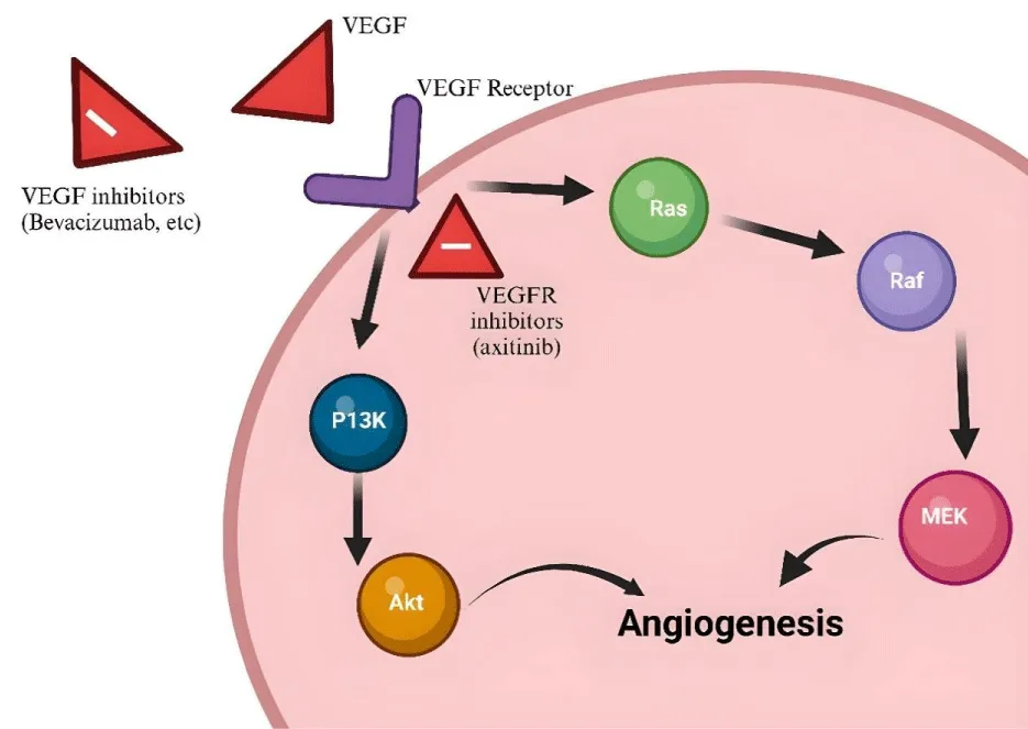
Figure 7: VEGFR activation and signal transduction.
Tumor angiogenesis and VEGFR-2 signaling represent critical processes in cancer progression. The tumor angiogenic pathways function in diverse mechanisms via sprouting angiogenesis, intussusceptive angiogenesis, vascular co-option, vascular mimicry, and glomeruloid angiogenesis, which are normally activated by varied angiogenic stimulators and their receptors intertwined to give rise to specialized signaling pathways [63,64].
VEGFR-2 is an important receptor that mediates tumor angiogenesis and has been estimated as an important therapeutic target. Numerous tumor types are observed for the widespread expression of vascular endothelial growth factor receptor 2 (VEGFR2), which regulates tumor angiogenesis. VEGFR-2 on cancer cells drives tumor angiogenesis to sustain tumor growth [65-67].
The hypoxic tumor microenvironment further upregulates VEGF expression through HIF1 activation, creating a pro-angiogenic feedback loop. Hypoxia is a common feature of solid tumors and develops because rapid growth outstrips oxygen supply and impaired blood flow due to the formation of abnormal vessels. Hypoxia can activate angiogenesis, thereby enhancing invasiveness and risk of metastasis, increasing survival of tumor cells, and suppressing anti-tumor immunity while hampering therapeutic response [68,69].
In pancreatic cancer, VEGF levels correlate with disease stage, tumor burden, and survival. Nevertheless, the efficacy of anti-VEGFR-2 therapies (tyrosine kinases, monoclonal antibodies) is limited due to the emergence of resistance mechanisms through the activation of other vascularization pathways. According to experts, the emergence of acquired resistance remains a major obstacle restricting the broad clinical success of molecular targeted therapies [70-72].
As a result of exposure to molecular targeted agents, VEGFR-2 expression gets induced, indicating the significance of VEGFR-2 signaling in molecular targeted therapy for cancer patients. Combination treatment of EGFR-TKIs and VEGFR-2 inhibitors may help overcome EGFR-TKI resistance. The importance of creating more selective and effective VEGFR-2 inhibitors is highlighted by these findings, as it may help overcome resistance [73].
Clinical validation and limitations of current VEGFR-2 inhibitors
More than a dozen approved drugs demonstrate the clinical validation of VEGFR-2 as a therapeutic target. The introduction of sorafenib in 2005 has seen approval of drugs targeting the VEGF/VEGFR pathway for roughly 20 solid tumor types, normally with combination therapy. The following compounds (2a-2d series) are multi-kinase inhibitors: Sunitinib, Pazopanib, Axitinib, Regorafenib, Cabozantinib, and Lenvatinib, as shown in the given figure (Figure 8) [74].
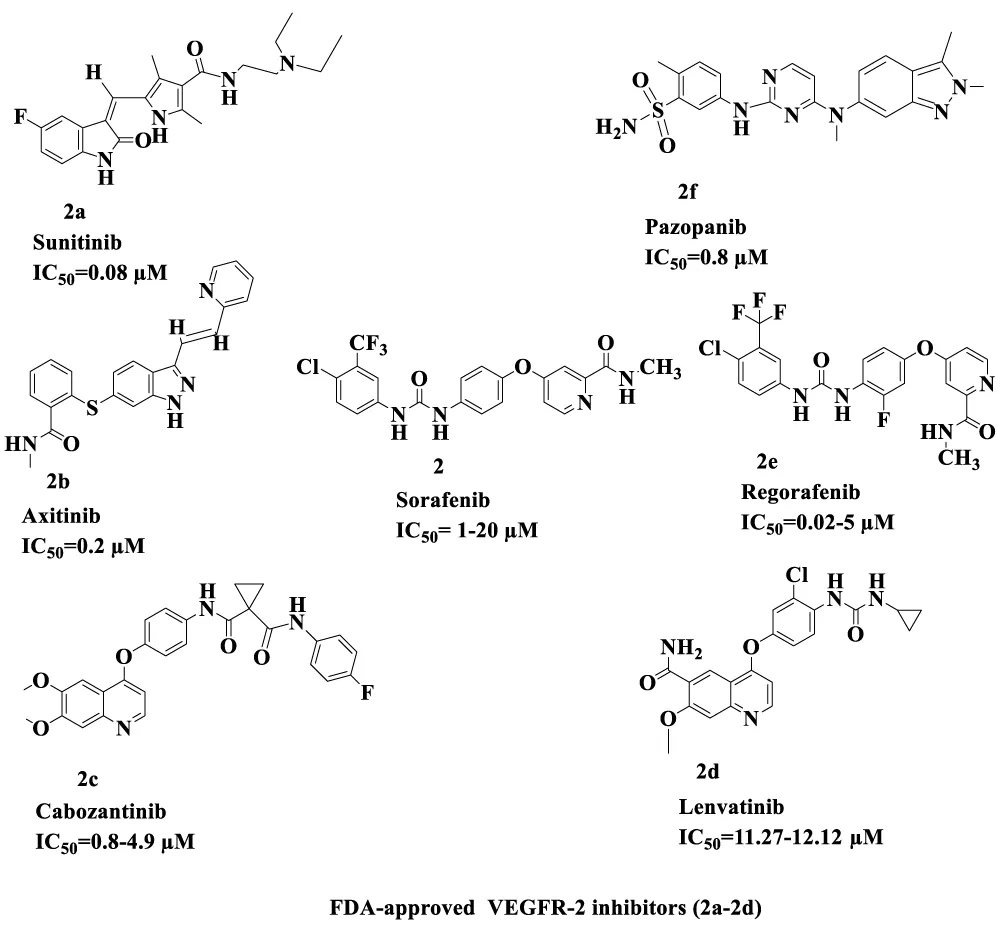
Figure 8: FDA-approved VEGFR-2 inhibitors (2a-2d).
Rivoceranib is a medication that inhibits a certain critical process in our body. The ANGEL study evaluated the effects of rivoceranib in patients with advanced or metastatic gastric or gastroesophageal junction cancer as a 3rd-line or 4th-line therapy. In patients treated with rivoceranib, PFS Progression-Free Survival), ORR(Objective Response Rate), and DCR Disease Control Rate) were all enhanced compared to placebo, and there was a prespecified 4th-line OS(Overall Survival) benefit as shown in the given (Figure 9) [75,76].
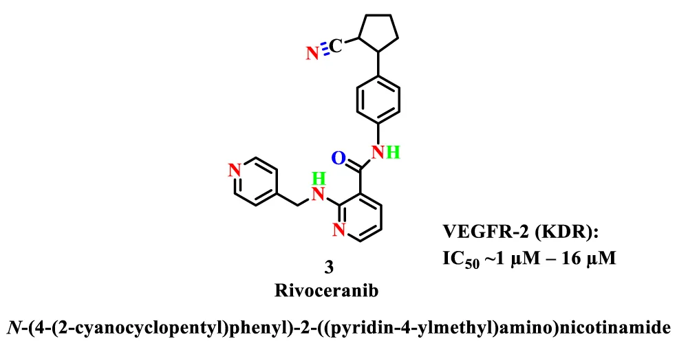
Figure 9: Structure of rivoceranib.
Despite clinical success, several challenges limit the therapeutic potential of current VEGFR-2 inhibitors. The widespread side effects linked to these VEGFR-2 inhibitors— hypertension, epistaxis, proteinuria, and upper respiratory infection—motivate researchers to search for new VEGFR-2 inhibitors with better pharmacokinetic profiles. Toxicities associated with available VEGFR-2 inhibitors are thought to be partly due to their effects against kinases other than VEGFR-2 [77,78].
The survival benefit has been modest in most tumor types, and there are currently no biomarkers in routine clinical use for identifying which patients are most likely to benefit from treatment. Nevertheless, the ability of these agents to reprogram the immunosuppressive tumor microenvironment into an immunostimulatory one [79,80].
Chemistry, synthesis, and structural diversity of 1,3,4-thiadiazole derivatives
Five-membered heterocyclic systems incorporating nitrogen along with oxygen or sulfur atoms have gained substantial attention in recent years as privileged scaffolds in the rational design of novel anticancer agents, as shown in the given figure (Figure 10). Within this class, thiazole derivatives have emerged as particularly attractive pharmacophores due to their diverse biological activities. Recent literature is beginning to point out the important antitumor potential of compounds based on thiadiazole, with particular focus on compounds that have a single, unfused, 2,5-disubstituted thiadiazole ring. A number of these derivatives have shown significant cytotoxic effects, with some being even more active than known reference anticancer drugs in preclinical studies. Part of the explanation of the strong therapeutic utility of the thiazole nucleus is to be found in its Bioisosteric similarity to pyrimidine, a structural motif which occurs in three of the five DNA and RNA nucleobases. Due to this structural similarity, thiazole derivatives have the potential to bind to nucleic acid-related biological processes, and thus, they may interfere with DNA replication and other cellular activities that are critical to the cellular growth of cancer. This property highlights why thiazoles are promising molecular scaffolds in the creation of second-generation anticancer therapeutics [81-83].
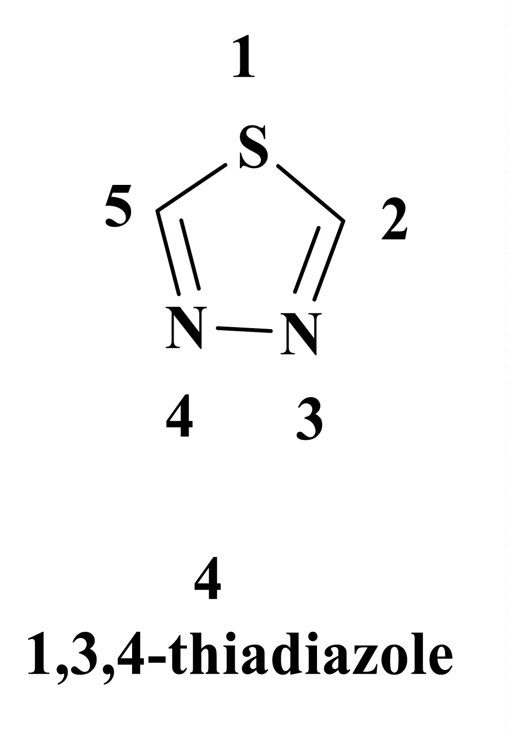
Figure 10: Structure of 1,3,4-thiadiazole.
Structurally, the thiazole nucleus is known to exist in various isomeric forms depending on the relative orientation of the nitrogen and sulfur heteroatoms in the five-membered ring. According to these position changes, there are four different isomeric forms of thiadiazole, namely 1,2,3-thiadiazole, 1,2,4-thiadiazole, 1,2,5-thiadiazole, and 1,3,4-thiadiazole, as shown in the following (Figure 11). The isomers have different electronic and steric properties, which may have a considerable impact on target selectivity and biological performance [84].
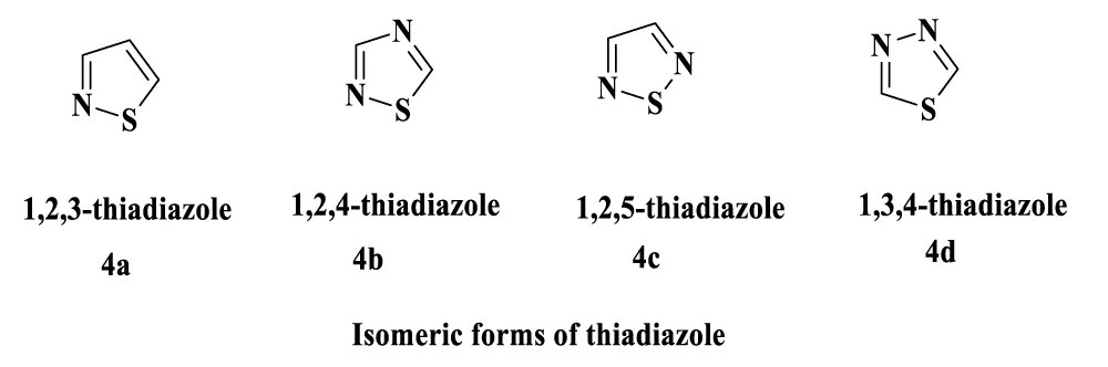
Figure 11: Distinct isomeric forms of thiadiazole.
Such a wide range of diverse biological activities of thiazole derivatives can be explained by the fact that they have a high capacity to interact with a variety of biological targets via a broad range of non-covalent and coordination-based interactions. These are hydrogen bonding, van der Waals interactions, hydrophobic forces, as well as metal ion coordination that work together to make them bind together, as well as their pharmacological versatility. This type of interactional versatility allows thiazole scaffolds to interact well with enzymes, receptors, and nucleic acid-related targets [85,86].
General synthetic methodologies and strategies
1,3,4-thiadiazole derivatives can be synthesized in a variety of ways, such as cyclization of linear organic derivatives. Various synthetic methods have been designed, and each has its benefits in terms of yields, reaction conditions, and the availability of starting materials [87,88].
Cyclization of Acylthiosemicarbazides: This is done by reacting acylthiosemicarbazides with dehydrating reagents, including phosphorus oxychloride, concentrated sulfuric acid, or polyphosphoric acid, in the presence of heating conditions. The process of cyclization is carried out by the nucleophilic attack of the terminal nitrogen on the carbon atom of the carbonyl group, and then the removal of water to produce the thiadiazol ring [89].
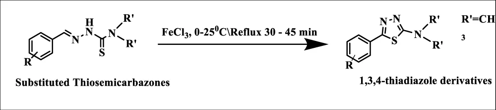
Oxidative Cyclization of Thiosemicarbazones: Oxidative cyclization of thiosemicarbazones can be performed using reagents like ferric chloride, iodine, or oxidizing agents to form 1,3,4-thiadiazoles. The technique has the advantage of mild reaction conditions and the ability to tolerate different functional groups. [90].
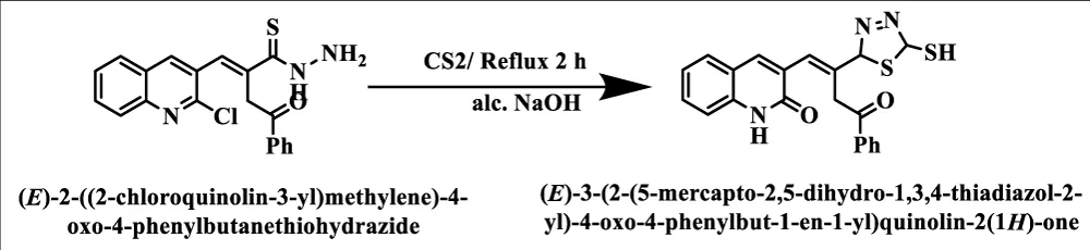
Reaction of Hydrazides with Carbon Disulfide: Acyl or acid hydrazide reacts with carbon disulfide in the presence of a base (usually potassium hydroxide) and produces 2-mercapto1,3,4- thiadiazole. These thiol derivatives are very versatile intermediates that can be further functionalized by alkylation, acylation, or even oxidation reactions [91].
Green Chemistry Approaches: Modern synthetic strategies include the synthesis catalyzed by microwaves, solvent-free conditions, and recyclable catalytic systems that are recyclable. [86]. These techniques reduce the reaction time, yield higher yields, and are less polluting. An example is the naphthamide derivatives that were made using microwave-assisted synthesis in a four-step procedure to produce compounds that have strong VEGFR-2 inhibitory effects [92,93].
Molecular hybridization and multi-pharmacophore approaches
Molecular hybridization, combining two or more pharmacophoric units into a single molecular entity, has emerged as a powerful strategy for developing improved therapeutic agents. This approach leverages the complementary properties of different scaffolds to achieve synergistic effects, multi-target inhibition, or improved pharmacokinetic profiles [94,95].
New benzothiazole hybrids linked to thiadiazole moieties have been synthesized and evaluated as VEGFR-2 inhibitors. A new series 5a-5d of 2-aminobenzothiazole hybrids linked to 1,3,4-thiadiazole aryl urea moieties was synthesized as shown in Figure 12, with compounds 5a-5d series showing a strong impact on cancer cell lines. The most promising compounds exhibited nanomolar VEGFR-2 inhibition, demonstrating the potential of combining thiazole with other privileged scaffolds [96-98].
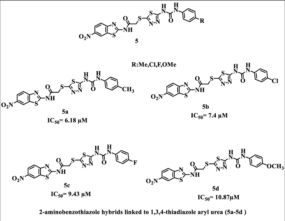
Figure 12: 2-aminobenzothiazole hybrids linked to 1,3,4-thiadiazole aryl urea moieties (5a-5d).
Novel quinazolines bearing 1,3,4-thiadiazole-aryl urea derivatives were designed as anticancer agents. A novel series 6a-6d of 1-(5-((6-nitroquinazoline-4-yl) thio)-1,3,4-thiadiazol-2yl)-3-phenylurea derivatives was synthesized to evaluate their cytotoxic potencies against multiple cancer cell lines, as shown in the given (Figure 13). Computational investigations, including molecular dynamics, frontier molecular orbital analysis, Fukui reactivity descriptors, and electrostatic potential surface studies, were performed to illustrate structure-activity relationships [99,100].
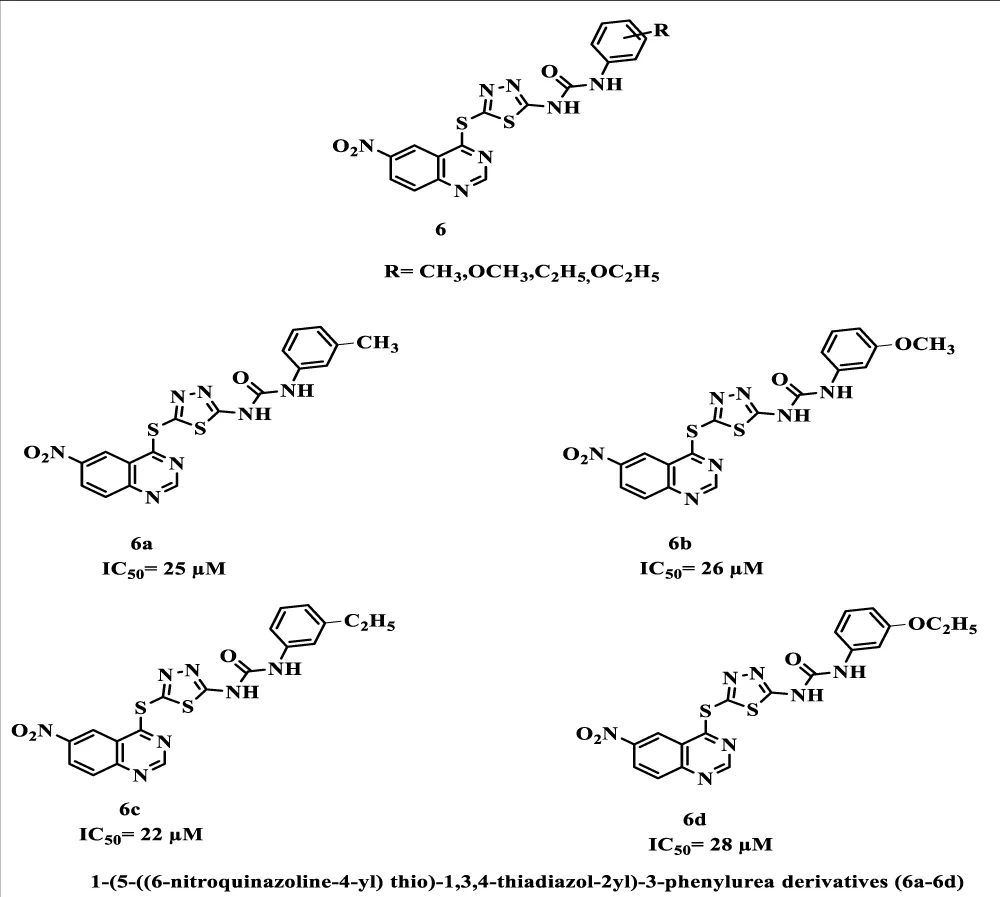
Figure 13: 1-(5-((6-nitroquinazoline-4-yl) thio)-1,3,4-thiadiazol-2yl)-3-phenylurea derivatives (6a-6d).
Expedition of sulfur-containing heterocyclic derivatives as cytotoxic agents in medicinal chemistry reveals that sulfur heterocyclic frameworks represent fundamental structures of diverse synthetic analogs with myriad therapeutic activities [101,102]. The incorporation of five and six-membered sulfur-containing scaffolds such as thiazoles, thiadiazoles, thiazolidinediones, and others has unveiled their effects through multiple mechanisms, including inhibition of tyrosine kinases, topoisomerase, tubulin, COX, DNA synthesis, and PI3K/Akt and Raf/MEK/ERK signaling pathways, as shown in the given (Figure 14) [103,104].
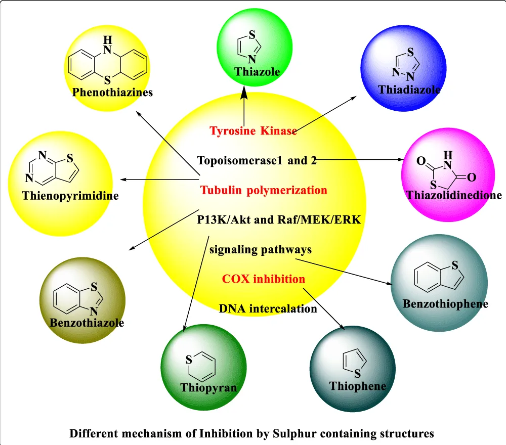
Figure 14: Different mechanisms of Inhibition by sulphur-containing structures.
The selectivity index (SI) serves as one of the most informative parameters in evaluating whether a newly synthesized compound preferentially targets cancer cells over healthy tissue. In the studies reviewed here, SI is derived from the ratio of the cytotoxic concentration causing 50% reduction in normal cell viability (CC₅₀) to the half-maximal inhibitory concentration against the cancer cell line (IC₅₀), expressed as SI = CC₅₀ (normal cells) / IC₅₀ (cancer cells) [105]. A compound yielding a high SI value is therefore one that kills cancer cells at concentrations far below those harmful to normal tissue — a distinction of considerable practical importance in drug development. Cell viability in both normal and cancerous populations is routinely assessed using the MTT assay, a well-established colorimetric method based on mitochondrial reduction of 3-(4,5-dimethylthiazol-2-yl)-2,5-diphenyltetrazolium bromide [106]. Across the studies cited in this review, assay conditions are broadly consistent: cells are seeded at approximately 5×10³ per well in 96-well plates, exposed to serial dilutions of the test compound (typically ranging from 0.001 to 100 µM) for 48 hours at 37°C under a humidified atmosphere containing 5% CO₂, after which MTT reagent is added and absorbance measured. Sorafenib, with a VEGFR-2 inhibitory IC₅₀ of approximately 0.041 µM, is consistently used as the positive reference control, providing a meaningful benchmark against which the potency of novel thiazole derivatives can be judged. For normal cell cytotoxicity assessment, the choice of cell line varies by study but commonly includes WI-38 human embryonic lung fibroblasts, HDF human dermal fibroblasts, WISH human amnion epithelial cells, or MCF-10A non-tumorigenic breast epithelial cells [107]. As a general interpretive threshold, an SI greater than 2 is taken as evidence of meaningful selectivity toward cancer cells, while values exceeding 10 reflect a particularly wide therapeutic window. Where SI values are reported across the compounds discussed in this review, they are evaluated against these criteria to give an honest picture of each compound’s safety profile relative to its antiproliferative potency.
Recent advances: Thiadiazole-based VEGFR-2 inhibitors (2020-2025) 2,3-dihydro-1,3,4-thiadiazole derivatives as potent VEGFR-2 inhibitors
The reduced form of the thiadiazole scaffold, 2,3-dihydro-1,3,4-thiadiazole, has shown exceptional promise as a core structure for VEGFR-2 inhibitor development. Discovery of new thiadiazole-based VEGFR-2 inhibitors through rational design, synthesis, cytotoxicity assessment, and apoptosis induction studies has yielded highly promising candidates [102,108-110].
The approved inhibitor sorafenib and others targeting vascular endothelial growth factor receptor-2 (VEGFR-2) have had off-target toxicities and resistance issues. As already established, the drug target is involved in cancer therapy. This study aimed at the design and development of novel thiazole-based vascular endothelial growth factor receptor 2 inhibitors that are more selective and safer. A series (7a-7e )of 2,3-dihydro-1,3,4-thiadiazole compounds was designed and synthesized [111].
In vitro cytotoxicity was determined against MCF-7, HepG-2, HCT-116, and normal WI-38 cells. Compound 7b was the most potent among the series of 7a-7e and selective with IC₅₀ values of 9.49 µM for MCF-7 and 12.89 µM for HepG-2 and more than three selectivity indices. The compound caused over 70% apoptosis and dual-phase (S and G2/M) cell cycle arrest, as shown in Figure 15 [112].
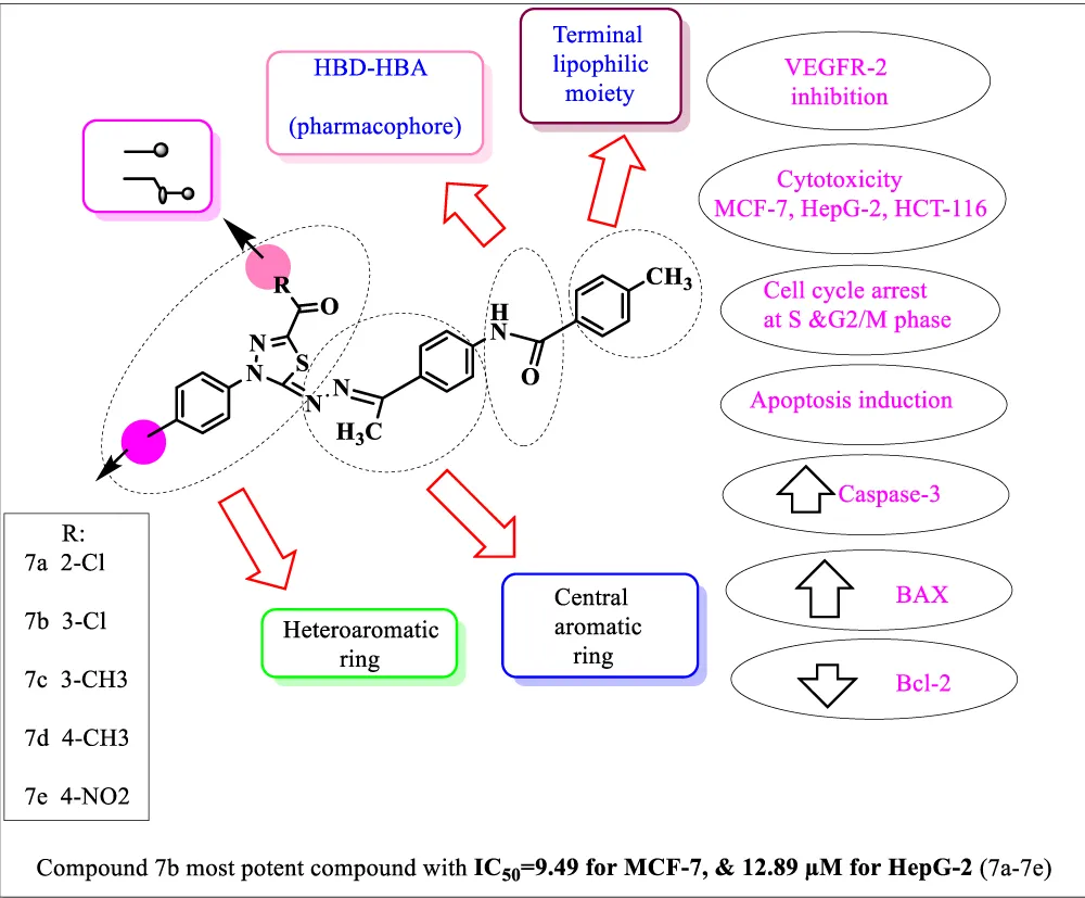
Figure 15: 2,3-dihydro-1,3,4-thiadiazole (7a-7e).
VEGFR-2 inhibition was demonstrated with IC₅₀ = 0.055 µM, comparable to sorafenib. Molecular docking, 200-ns molecular dynamics simulations, density functional theory calculations, and in silico toxicity profiling supported experimental findings. Computational studies confirmed stable binding at VEGFR-2 active sites, and compound 7b is a promising thiazole-based candidate with notable in vitro potency, selectivity, and mechanistic activity [113].
The anticancer, apoptotic, and VEGFR-2 inhibitory design and synthesis of thiadiazoles led to the discovery of remarkably active compounds. VEGFR-2 inhibitors are important in the treatment of cancer as they inhibit tumor angiogenesis. The synthesized compounds were screened for antiproliferative activity against human cancer cell lines (HCT-116, MCF-7, and HepG-2) and WI-38 as normal cells using sorafenib as a reference drug [114].
The compound 8(4,5-dihydro-1,3,4-thiadiazole) was the most potent anti-proliferative compound tested and displayed strong VEGFR-2 inhibition with an IC₅₀ of 41.51 µM, more potent than sorafenib, IC₅₀ =53.32 µM. The work showed that 8 slowed MCF-7 cells in their cell cycle analysis at the G2 phase. The low level of apoptosis (2%) was increased with an IC₅₀ of 700 µM after 48 h incubation, 53%, and associated with a greater than 12-fold increase in Bax/Bcl-2 ratio and activation of caspase-8/9. Moreover, 8 decreased the wound closure of MCF-7 cells to 5.28%, indicating strong anti-metastatic properties shown in the given (Figure 16) [115].
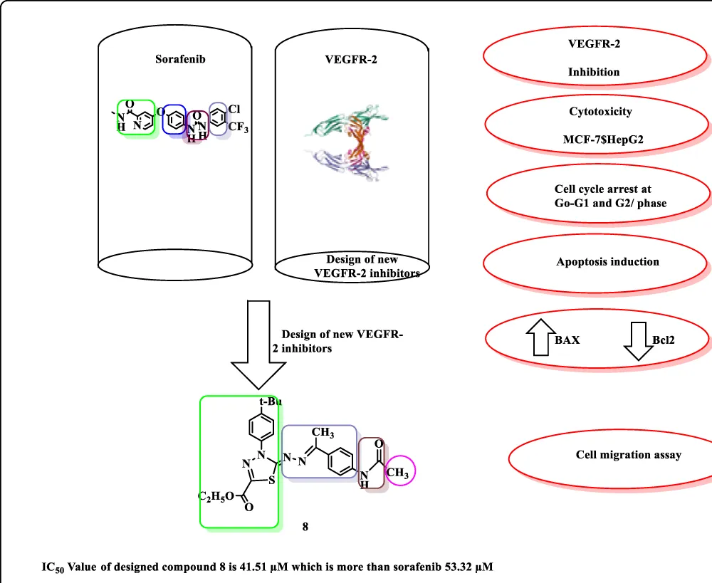
Figure 16: 4,5-dihydro-1,3,4-thiadiazole compound Promising Activity.
Dual BRaf/VEGFR-2 inhibitory thiadiazoles
The development of dual-target inhibitors represents an advanced strategy in cancer therapy, addressing multiple oncogenic pathways simultaneously. New 1,3,4-thiadiazole-based dual BRaf/VEGFR-2 inhibitors with potential anti-breast cancer activity have been developed. This study reported the design, synthesis, and biological evaluation of a novel series 9a-9f of 1,3,4-thiadiazole-based derivatives as dual BRaf/VEGFR-2 kinase inhibitors with potential anticancer activity. Among the whole series 9a-9f synthesized compounds, 9b emerged as the most potent candidate, exhibiting strong cytotoxicity against MDA-MB-231 and MCF-7 breast cancer cell lines (IC₅₀ = 9.66 and 15.83 µM, respectively), with minimal toxicity toward normal WI-38 and WISH cells, reflected by favorable selectivity indices as shown in Figure 17. Compound 9b, featuring a unique structural assembly of a 2,3-dihydro-1,3,4-thiadiazole core, para-methoxyphenyl group, and sulfonamide-linked methylpiperidine moiety, exhibited superior dual-inhibitory activity with IC₅₀ values of 0.75 µM for BRaf and 58.13 µM for VEGFR-2. Flow cytometry and gene expression analysis revealed that the compound induces G1-phase cell cycle arrest and promotes apoptosis through upregulation of BAX and caspases-8/9, with downregulation of Bcl-2. Structure-activity relationship analysis indicated that substitution with electron-donating groups enhanced cytotoxic potency. Molecular docking and molecular dynamics simulations confirmed stable binding to active sites, supported by Glide scores (-25.47 and -31.64 kcal/mol) and MMGBSA energies, while DFT(Density Functional Theory) calculations further validated the compound’s electronic stability and reactivity [116].
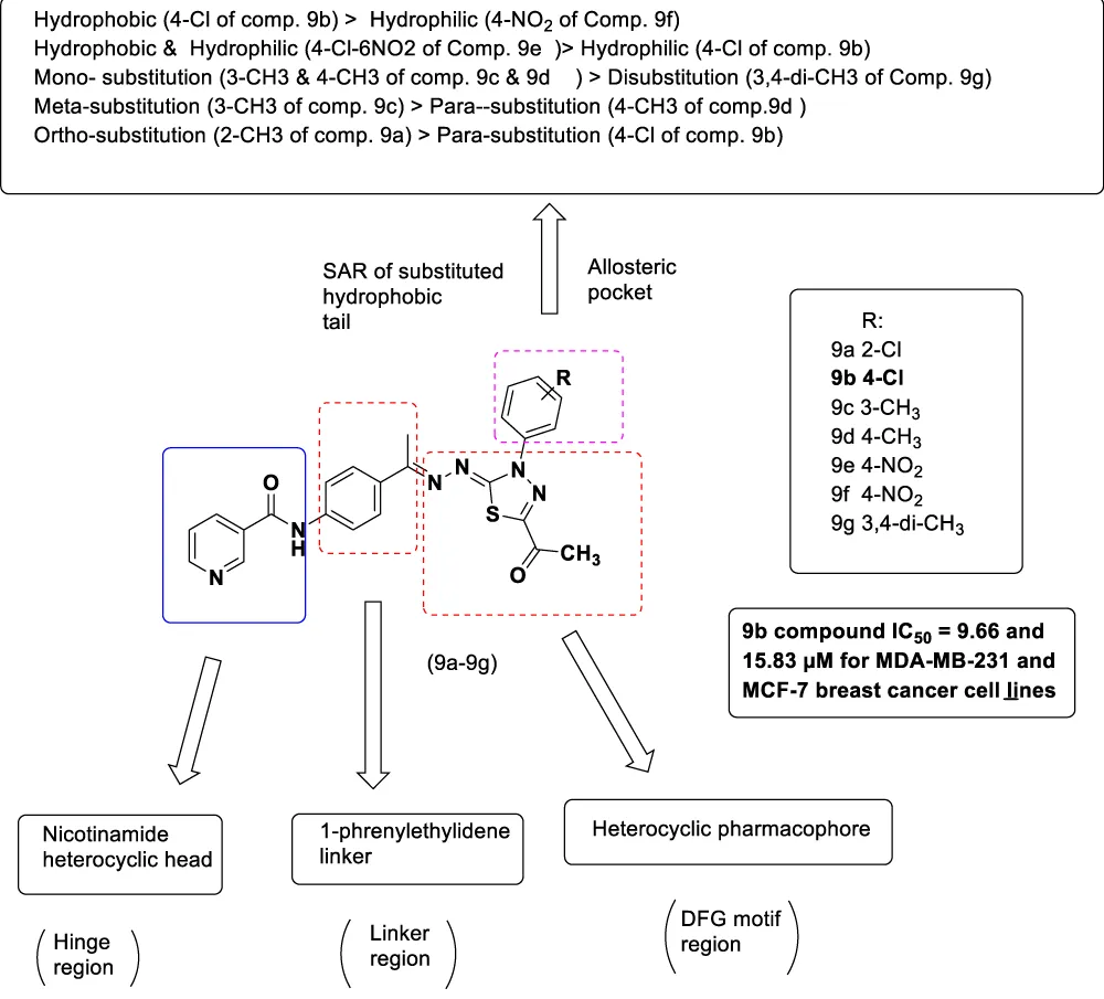
Figure 17: New 1,3,4-thiadiazole based dual BRaf/VEGFR-2 inhibitors (9a-9g).
Identification of benzothiazoles bearing 1,3,4-thiadiazole as antiproliferative hybrids targeting VEGFR-2 and BRAF kinase yielded compounds (10a-10f) with exceptional properties, demonstrating remarkable cytotoxicity with IC₅₀ values ranging from 3.58 to 15.36 µM against three cancer cell lines, showing IC₅₀ values of 38.77-66.22 µM against normal cell lines, significantly safer than sorafenib, as shown in Figure 18. Compound 10f exhibited the capacity to Inhibit both BRAF and VEGFR-2 enzymes with IC₅₀ values similar to sorafenib (0.071 and 0.194 µM, respectively) and caused G2-M- and S-phase cycle arrest as shown in Figure 18 [117].
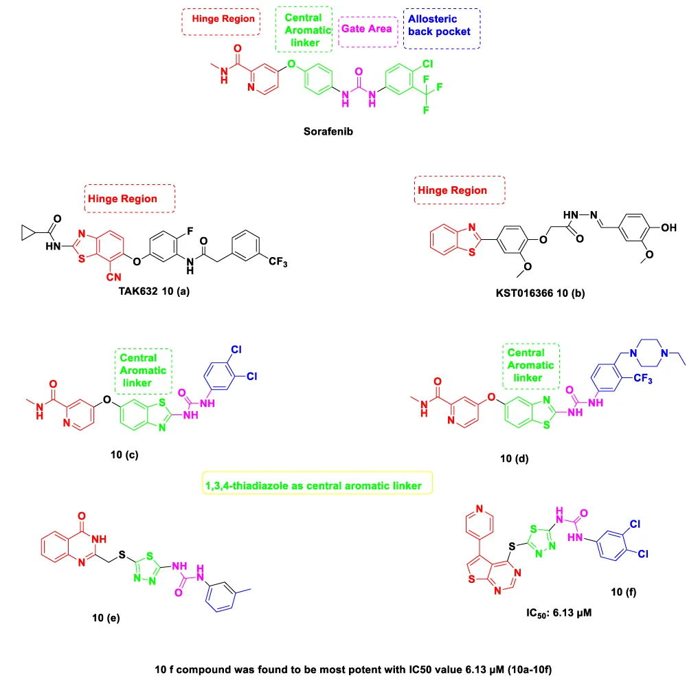
Figure 18: Benzothiazoles bearing 1,3,4-thiadiazole (10a-10f).
The new thiazole derivatives produced as VEGFR-2 inhibitors with anticancer and proapoptotic activities, out of which compound 10f was produced, which demonstrated cytotoxic activity against MCF7 breast cancer cells (IC₅₀: 6.13 µM) higher than that of sorafenib (IC₅₀: 7.26 µM). The compound has potent VEGFR-2 inhibition (IC₅₀: 40.65 µM), more potent than sorafenib (IC₅₀: 53.32 µM). Apoptosis analysis revealed primarily late apoptosis or necrosis (21.81%), with treatment increasing expression of proapoptotic gene BAX (4.19 ± 0.34-fold) and suppressing antiapoptotic Bcl-2 (0.38 ± 0.02-fold), resulting in a dramatic 11.03-fold increase in BAX/Bcl-2 ratio.Caspase-8 and caspase-9 levels were elevated by 2.99- and 4.13-fold, respectively, confirming activation of both intrinsic and extrinsic apoptotic pathways [118-120].
Different biological activities of thiadiazole
1,3,4-Thiadiazole is a five-membered aromatic heterocycle containing two nitrogen atoms and one sulfur atom positioned at the 1, 3, and 4 positions of the ring. The thiazole scaffold is widely studied for its diverse pharmacological activities, exhibiting bioactivity as an anticancer agent toward human cancers, along with antibacterial, diuretic, antitubercular, and antifungal properties, as shown below (Figure 19) [117,121-123].
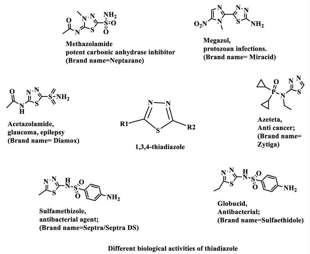
Figure 19: Different activities of thiazole.
The unique pharmacological profile arises from distinctive electronic properties. The thiazole ring consists of sulfur and nitrogen in such a fashion that electrons are free to move from one bond to another, rendering aromatic properties. On account of its aromaticity, reactive positions exist where donor-acceptor, nucleophilic, and oxidation reactions may take place [124-127].
The mesoionic character of the 1,3,4-thiadiazole ring involves delocalization of electrons across the heterocyclic system, with the sulfur atom playing a crucial role in electron distribution. The N-N-C-S motif and sulfur atom of thiadiazole significantly contribute to VEGFR-2 binding through key molecular interactions. The nitrogen atoms, with their lone pairs of electrons, serve as effective hydrogen bond acceptors, facilitating interactions with biological macromolecules [128,129].
The heteroatom used to form the thiadiazole ring is based on a benzene ring or a simple aromatic. Thiadiazole derivatives have been known to display diverse pharmacological activity, and several of their related compounds have multiple biologically active constituents and various applications that have been clinically approved. In recent years, several biologically active thiazole and bisthiazole derivatives have been reported in the literature due to their great medicinal importance [130-132].
The overall discussion of thiazole-based VEGFR-2 inhibitors that have been developed in the last five years shows conclusively that the 1,3,4-thiazole scaffold forms a privileged heterocycle that can be used to come up with selective, potent, and safe anticancer agents. The systematic screening of more than 151 thiazole analogs demonstrates some regular trends in nanomolar to sub-micromolar VEGFR-2 inhibitory potentials, strong cytotoxic action in a variety of cancer cell lines, and desirable selectivity indices that can differentiate between cancer and normal cells.
The existence of critical molecular determinants such as the essential N-N-C-S motif, optimal linker lengths (3-5 bonds), the significance of electron-donating substituents, and the strategic positioning of halogen atoms has been detected by studies of structure-activity relationships.
Consistent mechanistic evidence indicates that VEGFR-2 inhibitors that contain thiadiazole will induce apoptosis by activating both intrinsic and extrinsic pathways, disrupt cell cycle progression at numerous points, and prevent cancer cell migration and invasion. The multi-mechanistic profile observed across these thiazole derivatives, taken together with their generally favorable in vitro selectivity relative to established agents like sorafenib, makes a reasonable case for their continued investigation as VEGFR-2-targeted anticancer candidates. That said, it would be premature to draw firm conclusions from the current body of evidence alone. Virtually all of the data discussed in this review originates from cell-based in vitro experiments, and while such findings are a necessary and valuable first step, they cannot by themselves reliably predict how a compound will behave in a living biological system. Factors such as metabolic stability, tissue distribution, off-target organ toxicity, and pharmacokinetic behavior — none of which are adequately captured by cell culture models — will ultimately determine whether the promising selectivity profiles reported here translate into genuine therapeutic benefit. Any comparison with sorafenib or other clinically approved agents in terms of safety or efficacy must therefore be treated cautiously at this stage, as the absence of head-to-head in vivo data leaves such comparisons necessarily incomplete. Moving forward, it will be important for researchers working in this area to bridge this gap by subjecting the most promising lead compounds to rigorous animal model studies, encompassing both efficacy assessment in tumor-bearing models and thorough pharmacokinetic and toxicological profiling. Only through such validation can the current in vitro promise of thiazole-based VEGFR-2 inhibitors be meaningfully translated into preclinical candidates worthy of further advancement.
The combination of medicinal chemistry, structural biology, computational modeling, and systems biology methods has enabled the discovery and optimization of thiazoles against the mitigation of VEGFR-2. With the continued development of studies, the compounds may prove to be of great benefit to cancer patients. They may be more selective, less toxic, and have the potential to overcome resistance mechanisms of therapeutic agents currently in use. Thiadiazole-based derivatives are supported by strong evidence according to the review. VEGFR-2 inhibitors are a promising class of next-generation anticancer agents worthy of further investment and clinical translation.
The authors want to acknowledge with a deep sense of gratitude the faculty of pharmacy, Integral University, Lucknow, Uttar Pradesh, India, for the conducive academic ambience, necessary infrastructural facilities, and constant encouragement provided, which ultimately led to the successful completion of this review work.
Special thanks are given to the Department of Pharmaceutical Chemistry for allowing access to its institutional resources, including laboratory facilities, digital libraries, and scientific databases essential to conducting an extensive literature survey and compiling relevant data. I would also like to acknowledge the project sanction No: IUL/ICEIR/SMP/2024-08. The authors further extend their appreciation for the support of the staff of the university’s central library in facilitating access to peer-reviewed journals, books, and online repositories that were essential in this review.
The authors acknowledge with gratitude all those researchers and scientists whose original research findings were the basis of this review article. Their major contributions in medicinal chemistry, heterocyclic chemistry, and anticancer drug discovery have advanced scientific knowledge significantly and have led to this extensive analysis. They also want to thank other colleagues who have been helpful in the discussion and have supported them in the preparation of the manuscript. The authors finally wish to thank members of their family for being very supportive in their academic pursuits.
- Bray F, Laversanne M, Sung H, Ferlay J, Siegel RL, Soerjomataram I, et al. Global cancer statistics 2022: GLOBOCAN estimates of incidence and mortality worldwide for 36 cancers in 185 countries. CA Cancer J Clin. 2024 May;74(3):229–63. Available from: https://dx.doi.org/10.3322/caac.21834
- Zhang Y, Zhang M, Song H, Dai Q, Liu C. Tumor Microenvironment-Responsive Polymer-Based RNA Delivery Systems for Cancer Treatment. Small Methods. 2024 May;9(1). Available from: https://dx.doi.org/10.1002/smtd.202400278
- Bray F, Laversanne M, Weiderpass E, Soerjomataram I. The ever‐increasing importance of cancer as a leading cause of premature death worldwide. Cancer. 2021 Aug 15;127(16):3029–30. Available from: https://dx.doi.org/10.1002/cncr.33587
- Chen S, Cao Z, Prettner K, Kuhn M, Yang J, Jiao L, et al. Estimates and Projections of the Global Economic Cost of 29 Cancers in 204 Countries and Territories From 2020 to 2050. JAMA Oncol. 2023 Apr 1;9(4):465. Available from: https://dx.doi.org/10.1001/jamaoncol.2022.7826
- Nishida N, Yano H, Nishida T, Kamura T, Kojiro M. Angiogenesis in cancer. Vasc Health Risk Manag. 2006 Aug;2(3):213–9. Available from: https://dx.doi.org/10.2147/vhrm.2006.2.3.213
- Saman H, Raza SS, Uddin S, Rasul K. Inducing Angiogenesis, a Key Step in Cancer Vascularization, and Treatment Approaches. Cancers. 2020 May 6;12(5):1172. Available from: https://dx.doi.org/10.3390/cancers12051172
- Lee C, Kim MJ, Kumar A, Lee HW, Yang Y, Kim Y. Vascular endothelial growth factor signaling in health and disease: from molecular mechanisms to therapeutic perspectives. Signal Transduct Target Ther. 2025 May 19;10(1):170. Available from: https://dx.doi.org/10.1038/s41392-025-02249-0
- Mabeta P, Steenkamp V. The VEGF/VEGFR Axis Revisited: Implications for Cancer Therapy. Int J Mol Sci. 2022 Dec 9;23(24):15585. Available from: https://dx.doi.org/10.3390/ijms232415585
- Ribatti D, Annese T, Tamma R. Vascular co-option in resistance to anti-angiogenic therapy. Front Oncol. 2023 Dec 11;13:1323350. Available from: https://dx.doi.org/10.3389/fonc.2023.1323350
- Shah AA, Kamal MA, Akhtar S. Tumor Angiogenesis and VEGFR-2: Mechanism, Pathways and Current Biological Therapeutic Interventions. Curr Drug Metab. 2021 Mar 22;22(1):50–9. Available from: https://dx.doi.org/10.2174/1389200221666201019143252
- Marques CS, Brandão P, Burke AJ. Targeting Vascular Endothelial Growth Factor Receptor 2 (VEGFR-2): Latest Insights on Synthetic Strategies. Molecules. 2024 Nov 13;29(22):5341. Available from: https://dx.doi.org/10.3390/molecules29225341
- Shibuya M. Vascular Endothelial Growth Factor (VEGF) and Its Receptor (VEGFR) Signaling in Angiogenesis: A Crucial Target for Anti- and Pro-Angiogenic Therapies. Genes And Cancer. 2011 Oct;2(12):1097–105. Available from: https://dx.doi.org/10.1177/1947601911423031
- Ghalehbandi S, Yuzugulen J, Pranjol MZI, Pourgholami MH. The role of VEGF in cancer-induced angiogenesis and the research progress of drugs targeting VEGF. Eur J Pharmacol. 2023 Jun;949:175586. Available from: https://dx.doi.org/10.1016/j.ejphar.2023.175586
- Shibuya M. Vascular Endothelial Growth Factor (VEGF)-Receptor2: Its Biological Functions, Major Signaling Pathway, and Specific Ligand VEGF-E. Endothelium. 2006 Jan;13(2):63–9. Available from: https://dx.doi.org/10.1080/10623320600697955
- Shaik F, Cuthbert G, Homer-Vanniasinkam S, Muench S, Ponnambalam S, Harrison M. Structural Basis for Vascular Endothelial Growth Factor Receptor Activation and Implications for Disease Therapy. Biomolecules. 2020 Dec 15;10(12):1673. Available from: https://dx.doi.org/10.3390/biom10121673
- Risau W. Mechanisms of angiogenesis. Nature. 1997 Apr;386(6626):671–4. Available from: https://dx.doi.org/10.1038/386671a0
- Blondelle SE, Pérez-Payá E, Dooley CT, Pinilla C, Houghten RA. Soluble combinatorial libraries of organic, peptidomimetic, and peptide diversities. Trends Anal Chem. 1995 Feb;14(2):83–92. Available from: https://dx.doi.org/10.1016/0165-9936(95)91476-9
- Modi SJ, Kulkarni VM. Exploration of structural requirements for the inhibition of VEGFR-2 tyrosine kinase: Binding site analysis of type II, ‘DFG-out’ inhibitors. J Biomol Struct Dyn. 2022 Aug 13;40(12):5712–27. Available from: https://dx.doi.org/10.1080/07391102.2021.1872417
- Sanphanya K, Wattanapitayakul SK, Phowichit S, Fokin VV, Vajragupta O. Novel VEGFR-2 kinase inhibitors identified by the back-to-front approach. Bioorg Med Chem Lett. 2013 May;23(10):2962–7. Available from: https://dx.doi.org/10.1016/j.bmcl.2013.03.042
- Mahaki H, Nobari S, Tanzadehpanah H, Babaeizad A, Kazemzadeh G, Mehrabzadeh M, et al. Targeting VEGF signaling for tumor microenvironment remodeling and metastasis inhibition: Therapeutic strategies and insights. Biomed Pharmacother. 2025 May;186:118023. Available from: https://dx.doi.org/10.1016/j.biopha.2025.118023
- Buzatu IM, Tataranu LG, Duta C, Stoian I, Alexandru O, Dricu A. A Review of FDA-Approved Multi-Target Angiogenesis Drugs for Brain Tumor Therapy. Int J Mol Sci. 2025 Feb 28;26(5):2192. Available from: https://dx.doi.org/10.3390/ijms26052192
- Jang S, Strickland B, Finis L, Kooijman JJ, Melis JJTM, Zaman GJR, et al. Comparative biochemical kinase activity analysis identifies rivoceranib as a highly selective VEGFR2 inhibitor. Cancer Chemother Pharmacol. 2023 Jun;91(6):491–9. Available from: https://dx.doi.org/10.1007/s00280-023-04534-7
- Llovet JM, Ricci S, Mazzaferro V, Hilgard P, Gane E, Blanc JF, et al. Sorafenib in Advanced Hepatocellular Carcinoma. N Engl J Med. 2008 Jul 24;359(4):378–90. Available from: https://dx.doi.org/10.1056/NEJMoa0708857
- Motzer RJ, Hutson TE, Tomczak P, Michaelson MD, Bukowski RM, Rixe O, et al. Sunitinib versus Interferon Alfa in Metastatic Renal-Cell Carcinoma. N Engl J Med. 2007 Jan 11;356(2):115–24. Available from: https://dx.doi.org/10.1056/NEJMoa065044
- Rini BI, Escudier B, Tomczak P, Kaprin A, Szczylik C, Hutson TE, et al. Comparative effectiveness of axitinib versus sorafenib in advanced renal cell carcinoma (AXIS): a randomised phase 3 trial. The Lancet. 2011 Dec;378(9807):1931–9. Available from: https://dx.doi.org/10.1016/S0140-6736(11)61613-9
- Sternberg CN, Davis ID, Mardiak J, Szczylik C, Lee E, Wagstaff J, et al. Pazopanib in Locally Advanced or Metastatic Renal Cell Carcinoma: Results of a Randomized Phase III Trial. J Clin Oncol. 2010 Feb 20;28(6):1061–8. Available from: https://dx.doi.org/10.1200/JCO.2009.23.9764
- Choueiri TK, Escudier B, Powles T, Mainwaring PN, Rini BI, Donskov F, et al. Cabozantinib versus Everolimus in Advanced Renal-Cell Carcinoma. N Engl J Med. 2015 Nov 5;373(19):1814–23. Available from: https://dx.doi.org/10.1056/NEJMoa1510016
- Fuchs CS, Tomasek J, Yong CJ, Dumitru F, Passalacqua R, Goswami C, et al. Ramucirumab monotherapy for previously treated advanced gastric or gastro-oesophageal junction adenocarcinoma (REGARD): an international, randomised, multicentre, placebo-controlled, phase 3 trial. The Lancet. 2014 Jan;383(9911):31–9. Available from: https://dx.doi.org/10.1016/S0140-6736(13)61719-5
- Hossain M, Habib I, Singha K, Kumar A. FDA-approved heterocyclic molecules for cancer treatment: Synthesis, dosage, mechanism of action, and their adverse effect. Heliyon. 2024 Jan;10(1):e23172. Available from: https://dx.doi.org/10.1016/j.heliyon.2023.e23172
- Chaudhry F, Munir R, Malik N. N-Heterocycles as Privileged Scaffolds in FDA-Approved Different NMEs of 2021: A Review. Lett Org Chem. 2023 Apr;20(4):287–99. Available from: https://dx.doi.org/10.2174/1570178620666221026095145
- Elkady H, Elgammal WE, Khalifa MM, Mahdy HA, Ibrahim AS, Saad AY, et al. Development of New Thiadiazole Derivatives as VEGFR‐2 Inhibitors With Anticancer and Proapoptotic Activities. Arch Pharm (Weinheim). 2025 Jul;358(7):e70042. Available from: https://dx.doi.org/10.1002/ardp.70042
- Indelicato S, Bongiorno D, Mauro M, Cascioferro S. Recent Developments of 1,3,4-Thiadiazole Compounds as Anticancer Agents. Pharmaceuticals. 2025 Apr 16;18(4):580. Available from: https://dx.doi.org/10.3390/ph18040580
- Anthwal T, Paliwal S, Nain S. Diverse Biological Activities of 1,3,4-Thiadiazole Scaffold. Chemistry. 2022 Dec 6;4(4):1654–71. Available from: https://dx.doi.org/10.3390/chemistry4040107
- Kumar D, Kumar H, Kumar V, Deep A, Sharma A, Marwaha MG, et al. Mechanism-based approaches of 1,3,4-thiadiazole scaffolds as potent enzyme inhibitors for cytotoxicity and antiviral activity. Med Drug Discov. 2023 Feb;17:100150. Available from: https://dx.doi.org/10.1016/j.medidd.2022.100150
- Patel VM, Patel NB, Chan-Bacab MJ, Rivera G, Humal TR, Gamit AS. Synthesis and computational studies of 1,3,4-thiadiazole and benzothiazole clubbed benzimidazole analogous as anti-tubercular and anti-protozoal agents. J Mol Struct. 2025 Jan;1319:139326. Available from: https://dx.doi.org/10.1016/j.molstruc.2024.139326
- Alsfouk BA, Elgammal WE, Elkady H, Mahdy HA, Hassan SM, Husein DZ, et al. Development of new thiazole-based compounds targeting VEGFR-2: In vitro anticancer evaluation, mechanistic investigations, and in silico studies. J Mol Struct. 2025 Dec;1348:143544. Available from: https://dx.doi.org/10.1016/j.molstruc.2025.143544
- Gawande P, Matore BW, Murmu A, Kumar A, Jana S, Roy PP, et al. 1,3,4‐Thiadiazole Derivatives as VEGFR‐2 Inhibitors and Their Molecular Insight for Cancer Therapy. Chem. Biodivers. 2025 Aug 26;e01361. Available from: https://dx.doi.org/10.1002/cbdv.202501361
- Elgammal WE, Elkady H, Yousef RG, Eldehna WM, Husein DZ, Amin FG, et al. New nicotinamide–thiazole hybrids as VEGFR-2 inhibitors for breast cancer therapy: design, synthesis and in silico and in vitro evaluation. RSC Adv. 2025;15(18):14477–98. Available from: https://dx.doi.org/10.1039/D5RA01223F
- Amaya H, Tanigawa N, Lu C, Matsumura M, Shimomatsuya T, Horiuchi T, et al. Association of vascular endothelial growth factor expression with tumor angiogenesis, survival, and thymidine phosphorylase/platelet-derived endothelial cell growth factor expression in human colorectal cancer. Cancer Lett. 1997 Nov;119(2):227–35. Available from: https://dx.doi.org/10.1016/S0304-3835(97)00280-2
- Hubbard SR, Miller WT. Receptor tyrosine kinases: mechanisms of activation and signaling. Curr Opin Cell Biol. 2007 Apr;19(2):117–23. Available from: https://dx.doi.org/10.1016/j.ceb.2007.02.010
- Shah FH, Nam YS, Bang JY, Hwang IS, Kim DH, Ki M, et al. Targeting vascular endothelial growth receptor-2 (VEGFR-2): structural biology, functional insights, and therapeutic resistance. Arch Pharm Res. 2025 May;48(5):404–25. Available from: https://dx.doi.org/10.1007/s12272-025-01545-1
- Abdullah SE, Perez‐Soler R. Mechanisms of resistance to vascular endothelial growth factor blockade. Cancer. 2012 Jul 15;118(14):3455–67. Available from: https://dx.doi.org/10.1002/cncr.26540
- Abhinand CS, Raju R, Soumya SJ, Arya PS, Sudhakaran PR. VEGF-A/VEGFR2 signaling network in endothelial cells relevant to angiogenesis. J Cell Commun Signal. 2016 Dec;10(4):347–54. Available from: https://dx.doi.org/10.1007/s12079-016-0352-8
- Ahmad A, Nawaz MI. Molecular mechanism of VEGF and its role in pathological angiogenesis. J Cell Biochem. 2022 Dec;123(12):1938–65. Available from: https://dx.doi.org/10.1002/jcb.30344
- Arora A, Kivelä AM, Wang L, Minkeviciene R, Taskinen JH, Zhang B, et al. Protrudin regulates FAK activation, endothelial cell migration, and angiogenesis. Cell Mol Life Sci. 2022 Apr;79(4):220. Available from: https://dx.doi.org/10.1007/s00018-022-04251-z
- Blagoev B, Ong SE, Kratchmarova I, Mann M. Temporal analysis of phosphotyrosine-dependent signaling networks by quantitative proteomics. Nat Biotechnol. 2004 Sep;22(9):1139–45. Available from: https://dx.doi.org/10.1038/nbt1005
- Nkoana JK, More GK, Elhenawy AA, Mphahlele MJ. Examining the 2-aryl-5-nitrobenzofuran-based hydrazones for anti-breast (MCF-7) cancer activity, potential to induce cell cycle arrest, and inhibit receptor tyrosine kinases (VEGFR-2 & EGFR). Eur J Med Chem. 2025 Nov;298:118018. Available from: https://dx.doi.org/10.1016/j.ejmech.2025.118018
- Manning G, Whyte DB, Martinez R, Hunter T, Sudarsanam S. The Protein Kinase Complement of the Human Genome. Science. 2002 Dec 6;298(5600):1912–34. Available from: https://dx.doi.org/10.1126/science.1075762
- Vijayan RSK, He P, Modi V, Duong-Ly KC, Ma H, Peterson JR, et al. Conformational Analysis of the DFG-Out Kinase Motif and Biochemical Profiling of Structurally Validated Type II Inhibitors. J Med Chem. 2015 Jan 8;58(1):466–79. Available from: https://dx.doi.org/10.1021/jm501603h
- Weiss MM, Harmange JC, Polverino AJ, Bauer D, Berry L, Berry V, et al. Evaluation of a Series of Naphthamides as Potent, Orally Active Vascular Endothelial Growth Factor Receptor-2 Tyrosine Kinase Inhibitors. J Med Chem. 2008 Mar 1;51(6):1668–80. Available from: https://dx.doi.org/10.1021/jm701098w
- Folkman J. Role of angiogenesis in tumor growth and metastasis. Semin Oncol. 2002 Dec;29(6Q):15–8. Available from: https://dx.doi.org/10.1053/sonc.2002.37263
- Wang Z, Walker GW, Muir DC, Nagatani-Yoshida K. Toward a global understanding of chemical pollution: a first comprehensive analysis of national and regional chemical inventories. Environ Sci Technol. 2020;54(5):2575–84.
- Alitalo K, Carmeliet P. Molecular mechanisms of lymphangiogenesis in health and disease. Cancer Cell. 2002 Apr;1(3):219–27. Available from: https://dx.doi.org/10.1016/S1535-6108(02)00051-X
- Chaudhari PJ, Nemade AR, Shirkhedkar AA. Recent updates on the potential of VEGFR-2 small-molecule inhibitors as anticancer agents. RSC Adv. 2024;14(45):33384–417. Available from: https://dx.doi.org/10.1039/D4RA05244G
- Krishnamurty R, Maly DJ. Biochemical Mechanisms of Resistance to Small-Molecule Protein Kinase Inhibitors. ACS Chem Biol. 2010 Jan 15;5(1):121–38. Available from: https://dx.doi.org/10.1021/cb9002656
- Alkim C, Alkim H, Koksal AR, Boga S, Sen I. Angiogenesis in Inflammatory Bowel Disease. Int J Inflamm. 2015;2015:1–10. Available from: https://dx.doi.org/10.1155/2015/970890
- Benedito R, Rocha SF, Woeste M, Zamykal M, Radtke F, Casanovas O, et al. Notch-dependent VEGFR3 upregulation allows angiogenesis without VEGF–VEGFR2 signalling. Nature. 2012 Apr;484(7392):110–4. Available from: https://dx.doi.org/10.1038/nature10908
- Karami Fath M, Ebrahimi M, Nourbakhsh E, Zia Hazara A, Mirzaei A, Shafieyari S, et al. PI3K/Akt/mTOR signaling pathway in cancer stem cells. Pathol - Res Pract. 2022 Sep;237:154010. Available from: https://dx.doi.org/10.1016/j.prp.2022.154010
- Vara JÁF, Casado E, De Castro J, Cejas P, Belda-Iniesta C, González-Barón M. PI3K/Akt signalling pathway and cancer. Cancer Treat Rev. 2004 Apr;30(2):193–204. Available from: https://dx.doi.org/10.1016/j.ctrv.2003.07.007
- Peng Y, Wang Y, Zhou C, Mei W, Zeng C. PI3K/Akt/mTOR Pathway and Its Role in Cancer Therapeutics: Are We Making Headway? Front Oncol. 2022 Mar 24;12:819128. Available from: https://dx.doi.org/10.3389/fonc.2022.819128
- Lee BJ, Boyer JA, Burnett GL, Thottumkara AP, Tibrewal N, Wilson SL, et al. Selective inhibitors of mTORC1 activate 4EBP1 and suppress tumor growth. Nat Chem Biol. 2021 Oct;17(10):1065–74. Available from: https://dx.doi.org/10.1038/s41589-021-00813-7
- Li Z, Huang J, Wu J. pH-Sensitive nanogels for drug delivery in cancer therapy. Biomater Sci. 2021;9(3):574–89.
- Frentzas S, Lum C, Chen TY. Angiogenesis and Its Role in the Tumour Microenvironment: A Target for Cancer Therapy. In: Rajer M, Segelov E, editors. Current Cancer Treatment [Internet].. IntechOpen; 2020 [cited 2026 Jan 26].. Available from: https://www.intechopen.com/books/current-cancer-treatment/angiogenesis-and-its-role-in-the-tumour-microenvironment-a-target-for-cancer-therapy Available from: https://dx.doi.org/10.5772/intechopen.89667
- Liu Y, Kang C, Li M, Yu X, Liu H. Strategies and Progress of Raman Technologies for Cellular Uptake Analysis of the Drug Delivery Systems. Int J Nanomedicine. 2023 Nov;18:6883–900. Available from: https://dx.doi.org/10.2147/ijn.s435087
- Caldwell R, Bartoli M, Behzadian M, El-Remessy A, Al-Shabrawey M, Platt D, et al. Vascular Endothelial Growth Factor and Diabetic Retinopathy: Role of Oxidative Stress. Curr Drug Targets. 2005 Jun 1;6(4):511–24. Available from: https://dx.doi.org/10.2174/1389450054021981
- Chandler KB, Leon DR, Kuang J, Meyer RD, Rahimi N, Costello CE. N-Glycosylation regulates ligand-dependent activation and signaling of vascular endothelial growth factor receptor 2 (VEGFR2). J Biol Chem. 2019 Aug;294(35):13117–30. Available from: https://dx.doi.org/10.1074/jbc.RA119.008643
- Parkin DM, Bray F, Ferlay J, Pisani P. Global Cancer Statistics, 2002. CA Cancer J Clin. 2005 Mar 1;55(2):74–108. Available from: https://dx.doi.org/10.3322/canjclin.55.2.74
- Bray F, Ferlay J, Soerjomataram I, Siegel RL, Torre LA, Jemal A. Global cancer statistics 2018: GLOBOCAN estimates of incidence and mortality worldwide for 36 cancers in 185 countries. CA Cancer J Clin. 2018 Nov;68(6):394–424. Available from: https://dx.doi.org/10.3322/caac.21492
- Balkwill FR, Capasso M, Hagemann T. The tumor microenvironment at a glance. J Cell Sci. 2012 Dec 1;125(23):5591–6. Available from: https://dx.doi.org/10.1242/jcs.116392
- Grobbelaar C, Steenkamp V, Mabeta P. Vascular Endothelial Growth Factor Receptors in the Vascularization of Pancreatic Tumors: Implications for Prognosis and Therapy. Curr Issues Mol Biol. 2025 Mar 10;47(3):179. Available from: https://dx.doi.org/10.3390/cimb47030179
- Yeo D, Giardina C, Saxena P, Rasko JEJ. The next wave of cellular immunotherapies in pancreatic cancer. Mol Ther - Oncolytics. 2022 Mar;24:561–76. Available from: https://dx.doi.org/10.1016/j.omto.2022.01.010
- Kamisawa T, Wood LD, Itoi T, Takaori K. Pancreatic cancer. The Lancet. 2016 Jul;388(10039):73–85. Available from: https://dx.doi.org/10.1016/S0140-6736(16)00141-0
- Watanabe H, Ichihara E, Kayatani H, Makimoto G, Ninomiya K, Nishii K, et al. VEGFR2 blockade augments the effects of tyrosine kinase inhibitors by inhibiting angiogenesis and oncogenic signaling in oncogene‐driven non‐small‐cell lung cancers. Cancer Sci. 2021 May;112(5):1853–64. Available from: https://dx.doi.org/10.1111/cas.14801
- Star E, Stevens M, Gooding C, Smith CWJ, Li L, Ayine ML, et al. A drug-repositioning screen using splicing-sensitive fluorescent reporters identifies novel modulators of VEGF-A splicing with anti-angiogenic properties. Oncogenesis. 2021 May 3;10(5):36. Available from: https://dx.doi.org/10.1038/s41389-021-00323-0
- Kang YK, Ryu MH, Di Bartolomeo M, Chau I, Yoon H, Kim JG, et al. Rivoceranib, a VEGFR-2 inhibitor, monotherapy in previously treated patients with advanced or metastatic gastric or gastroesophageal junction cancer (ANGEL study): an international, randomized, placebo-controlled, phase 3 trial. Gastric Cancer. 2024 Mar;27(2):375–86. Available from: https://dx.doi.org/10.1007/s10120-023-01455-5
- Liu Q, Zhao Y, Tan Y, An Y, Zheng Y, Zheng C, et al. NanoRNP Overcomes Tumor Heterogeneity in Cancer Treatment. Nano Lett. 2019 Oct;19(11):7662–72. Available from: https://dx.doi.org/10.1021/acs.nanolett.9b02501
- Gordon EJ, Fukuhara D, Weström S, Padhan N, Sjöström EO, Van Meeteren L, et al. The endothelial adaptor molecule TSAd is required for VEGF-induced angiogenic sprouting through junctional c-Src activation. Sci Signal. 2016 Jul 19;9(437). Available from: https://dx.doi.org/10.1126/scisignal.aad9256
- Yogi A, Callera GE, Aranha AB, Antunes TT, Graham D, McBride M, et al. Sphingosine-1-Phosphate-Induced Inflammation Involves Receptor Tyrosine Kinase Transactivation in Vascular Cells: Upregulation in Hypertension. Hypertension. 2011 Apr;57(4):809–18. Available from: https://dx.doi.org/10.1161/HYPERTENSIONAHA.110.162719
- Zhao Y. Stem cells in gastric cancer. World J Gastroenterol. 2015;21(1):112. Available from: https://dx.doi.org/10.3748/wjg.v21.i1.112
- Ramjiawan RR, Griffioen AW, Duda DG. Anti-angiogenesis for cancer revisited: Is there a role for combinations with immunotherapy? Angiogenesis. 2017 May;20(2):185–204. Available from: https://dx.doi.org/10.1007/s10456-017-9552-y
- Szeliga M. Thiadiazole derivatives as anticancer agents. Pharmacol Rep. 2020 Oct;72(5):1079–100. Available from: https://dx.doi.org/10.1007/s43440-020-00154-7
- Ferlay J, Colombet M, Soerjomataram I, Mathers C, Parkin DM, Piñeros M, et al. Estimating the global cancer incidence and mortality in 2018: GLOBOCAN sources and methods. Int J Cancer. 2019 Apr 15;144(8):1941–53. Available from: https://dx.doi.org/10.1002/ijc.31937
- Haider S, Alam MS, Hamid H. 1,3,4-Thiadiazoles: A potent multi-targeted pharmacological scaffold. Eur J Med Chem. 2015 Mar;92:156–77. Available from: https://dx.doi.org/10.1016/j.ejmech.2014.12.035
- Stewart JA, Ackerly CC, Myers CF, Newman RA, Krakoff IH. Clinical and clinical pharmacologic studies of 2-amino-1,3,4-thiadiazole (A-TDA: NSC 4728). Cancer Chemother Pharmacol. 1986 Apr;16(3):287–91. Available from: https://dx.doi.org/10.1007/BF00293994
- Janowska S, Khylyuk D, Bielawska A, Szymanowska A, Gornowicz A, Bielawski K, et al. New 1,3,4-Thiadiazole Derivatives with Anticancer Activity. Molecules. 2022 Mar 10;27(6):1814. Available from: https://dx.doi.org/10.3390/molecules27061814
- Oubella A, Bimoussa A, Rehman MT, AlAjmi MF, Auhmani A, Taha ML, et al. Molecular hybrids based on 1,2,3-triazole and 1,3,4-thiadiazole cores: Synthesis, characterization, anticancer activity, and in silico study. J Mol Struct. 2024 Sep;1311:138339. Available from: https://dx.doi.org/10.1016/j.molstruc.2024.138339
- De Andrade Danin Barbosa G, Palermo De Aguiar A. Synthesis of 1,3,4-Thiadiazole Derivatives and Microbiological Activities: A Review. Rev Virtual Quím. 2019;11(3):806–48. Available from: https://dx.doi.org/10.21577/1984-6835.20190058
- Stecoza CE, Nitulescu GM, Draghici C, Caproiu MT, Hanganu A, Olaru OT, et al. Synthesis of 1,3,4-Thiadiazole Derivatives and Their Anticancer Evaluation. Int J Mol Sci. 2023 Dec 14;24(24):17476. Available from: https://dx.doi.org/10.3390/ijms242417476
- Singh AK, Karan J, Kundu A, Gogoi R, Bhatia A, Mondal K, et al. Virtual Screening-Based Synthesis of 1,3,4-Thiadiazole Derivatives and Their Evaluation for Fungicidal Activity, Ergosterol Inhibition, and Aquatic Toxicity. J Agric Food Chem. 2026 Jan 14;74(1):549–59. Available from: https://dx.doi.org/10.1021/acs.jafc.5c13886
- Singh SJ, Rajamanickam S, Gogoi A, Patel BK. Synthesis of 2-amino-substituted-1,3,4-thiadiazoles via 2,3-dichloro-5,6-dicyano-1,4-benzoquinone (DDQ) mediated intramolecular C–S bond formation in thiosemicarbazones. Tetrahedron Lett. 2016 Mar;57(9):1044–7. Available from: https://dx.doi.org/10.1016/j.tetlet.2016.01.083
- Makane VB, Krishna VS, Krishna EV, Shukla M, Mahizhaveni B, Misra S, et al. Novel 1,3,4-oxadiazoles as Antitubercular Agents with Limited Activity Against Drug-Resistant Tuberculosis. Future Med Chem. 2019 Mar;11(6):499–510. Available from: https://dx.doi.org/10.4155/fmc-2018-0378
- Dai P, Jiao J, Li Y, Teng P, Wang Q, Zhu Y, et al. Novel 5-Sulfonyl-1,3,4-thiadiazole-Substituted Flavonoids as Potential Bactericides and Fungicides: Design, Synthesis, Three-Dimensional Quantitative Structure–Activity Relationship Studies. J Agric Food Chem. 2024 Mar 27;72(12):6672–83. Available from: https://dx.doi.org/10.1021/acs.jafc.3c06367
- Wu W, Ji Y, Wang Z, Wu X, Li J, Gu F, et al. The FDA-approved anti-amyloid-β monoclonal antibodies for the treatment of Alzheimer’s disease: a systematic review and meta-analysis of randomized controlled trials. Eur J Med Res. 2023;28(1):544.
- Marchesi E, Perrone D, Navacchia ML. Molecular Hybridization as a Strategy for Developing Artemisinin-Derived Anticancer Candidates. Pharmaceutics. 2023 Aug 23;15(9):2185. Available from: https://dx.doi.org/10.3390/pharmaceutics15092185
- Marinescu M. Benzimidazole-Triazole Hybrids as Antimicrobial and Antiviral Agents: A Systematic Review. Antibiotics. 2023 Jul 22;12(7):1220. Available from: https://dx.doi.org/10.3390/antibiotics12071220
- Marinescu M. Synthesis of Antimicrobial Benzimidazole–Pyrazole Compounds and Their Biological Activities. Antibiotics. 2021 Aug 19;10(8):1002. Available from: https://dx.doi.org/10.3390/antibiotics10081002
- Hussein AM, EL‐Sofany WI, Alminderej FM, Albadri AEAE, Awad HM, Nossier ES, et al. New Thiadiazole‐Triazole Hybrid Glycosides as Potential EGFR and VEGFR‐2 Inhibitors: Synthesis, Anticancer Activity, and Docking Simulation. ChemistrySelect. 2025 Jul;10(28):e04887. Available from: https://dx.doi.org/10.1002/slct.202404887
- Al-Sanea MM, Hamdi A, Mohamed AAB, El-Shafey HW, Moustafa M, Elgazar AA, et al. New benzothiazole hybrids as potential VEGFR-2 inhibitors: design, synthesis, anticancer evaluation, and in silico study. J Enzyme Inhib Med Chem. 2023 Dec 31;38(1):2166036. Available from: https://dx.doi.org/10.1080/14756366.2023.2166036
- Wang Z, Shi XH, Wang J, Zhou T, Xu YZ, Huang TT, et al. Synthesis, structure–activity relationships and preliminary antitumor evaluation of benzothiazole-2-thiol derivatives as novel apoptosis inducers. Bioorg Med Chem Lett. 2011 Feb;21(4):1097–101. Available from: https://dx.doi.org/10.1016/j.bmcl.2010.12.124
- Masoudinia S, Samadizadeh M, Safavi M, Bijanzadeh HR, Foroumadi A. Novel quinazolines bearing 1,3,4-thiadiazole-aryl urea derivative as anticancer agents: design, synthesis, molecular docking, DFT and bioactivity evaluations. BMC Chem. 2024 Feb 12;18(1):30. Available from: https://dx.doi.org/10.1186/s13065-024-01119-0
- Maji S, Debnath B, Panda S, Manna T, Maity A, Dayaramani R, et al. Anticancer Potential of the S ‐Heterocyclic Ring Containing Drugs and Their Bioactivation to Reactive Metabolites. Chem. Biodivers. 2024 Jul;21(7):e202400473. Available from: https://dx.doi.org/10.1002/cbdv.202400473
- Laxmikeshav K, Kumari P, Shankaraiah N. Expedition of sulfur‐containing heterocyclic derivatives as cytotoxic agents in medicinal chemistry: A decade update. Med Res Rev. 2022 Jan;42(1):513–75. Available from: https://dx.doi.org/10.1002/med.21852
- Yousef RG, Eissa IH, Elkady H, M Mehany AB, Abo-Saif MA, Radwan MM, et al. Design and synthesis of new nicotinamides as immunomodulatory VEGFR-2 inhibitors and apoptosis inducers. Future Med Chem. 2024 Dec 16;16(24):2583–98. Available from: https://dx.doi.org/10.1080/17568919.2024.2421150
- Yousef RG, Elkady H, Elkaeed EB, Gobaara IMM, Al-ghulikah HA, Husein DZ, et al. (E)-N-(3-(1-(2-(4-(2,2,2-Trifluoroacetamido)benzoyl)hydrazono)ethyl)phenyl)nicotinamide: A Novel Pyridine Derivative for Inhibiting Vascular Endothelial Growth Factor Receptor-2: Synthesis, Computational, and Anticancer Studies. Molecules. 2022 Nov 9;27(22):7719. Available from: https://dx.doi.org/10.3390/molecules27227719
- Radha Abbas Hasoon M, Jawad Kadhim N. Improvement of the Selectivity Index (SI) and Cytotoxicity Activity of Doxorubicin Drug by Panax ginseng plant Extract. Arch Razi Inst. 2021 Aug;(Online First). Available from: https://dx.doi.org/10.22092/ari.2021.355413.1681
- Anand U, Dey A, Chandel AKS, Sanyal R, Mishra A, Pandey DK, et al. Cancer chemotherapy and beyond: Current status, drug candidates, associated risks and progress in targeted therapeutics. Genes Dis. 2023 Jul;10(4):1367–401. Available from: https://dx.doi.org/10.1016/j.gendis.2022.02.007
- Madhubala MM, Nayantara GS, Jayasree R, Locs J, Mahalaxmi S. Biogenic amorphous calcium phosphate: a sustainable alternative for dentin remineralization. BMC Oral Health. 2025 Jul 11;25(1):1149. Available from: https://dx.doi.org/10.1186/s12903-025-06524-y
- Toolabi M, Safari F, Ayati A, Fathi P, Moghimi S, Salarinejad S, et al. Synthesis of novel 2‐acetamide‐5‐phenylthio‐1,3,4‐thiadiazole‐containing phenyl urea derivatives as potential VEGFR‐2 inhibitors. Arch Pharm (Weinheim). 2022 Mar;355(3):2100397. Available from: https://dx.doi.org/10.1002/ardp.202100397
- Abbas HAS, Nossier ES, El-Manawaty MA, El-Bayaa MN. New sulfonamide-based glycosides incorporated 1,2,3-triazole as cytotoxic agents through VEGFR-2 and carbonic anhydrase inhibitory activity. Sci Rep. 2024 Jun 6;14(1):13028. Available from: https://dx.doi.org/10.1038/s41598-024-62864-9
- Eissa IH, Elgammal WE, Mahdy HA, Zara S, Carradori S, Husein DZ, et al. Design, synthesis, and evaluation of novel thiadiazole derivatives as potent VEGFR-2 inhibitors: a comprehensive in vitro and in silico study. RSC Adv. 2024;14(48):35505–19. Available from: https://dx.doi.org/10.1039/D4RA04158E
- Elkaeed EB, Elgammal WE, Elkady H, Mahdy HA, Alsfouk AA, Husein DZ, et al. Discovery of new thiadiazole-based VEGFR-2 inhibitors: design, synthesis, cytotoxicity, and apoptosis induction. Future Med Chem. 2025 Sep 2;17(17):2145–62. Available from: https://dx.doi.org/10.1080/17568919.2025.2552639
- Elsayed EA, Sharaf-Eldin MA, Wadaan M. In vitro Evaluation of Cytotoxic Activities of Essential Oil from Moringa oleifera Seeds on HeLa, HepG2, MCF-7, CACO-2, and L929 Cell Lines. Asian Pac J Cancer Prev. 2015 Jun 26;16(11):4671–5. Available from: https://dx.doi.org/10.7314/APJCP.2015.16.11.4671
- Elkaeed EB, Yousef RG, Elkady H, Mehany ABM, Alsfouk BA, Husein DZ, et al. In silico, in vitro VEGFR-2 inhibition, and anticancer activity of a 3-(hydrazonomethyl)naphthalene-2-ol derivative. J Biomol Struct Dyn. 2023 Nov 2;41(16):7986–8001. Available from: https://dx.doi.org/10.1080/07391102.2022.2127907
- Mahdy HA, Elkady H, Elgammal WE, Elkaeed EB, Alsfouk AA, Ibrahim IM, et al. Design, synthesis, in vitro, and in silico studies of new thiazole derivatives as promising VEGFR-2 inhibitors and apoptosis inducers. J Mol Struct. 2024 Nov;1316:139019. Available from: https://dx.doi.org/10.1016/j.molstruc.2024.139019
- Elkady H, Elgammal WE, Mahdy HA, Zara S, Carradori S, Husein DZ, et al. Anti-proliferative 2,3-dihydro-1,3,4-thiadiazoles targeting VEGFR-2: Design, synthesis, in vitro, and in silico studies. Comput Biol Chem. 2024 Dec;113:108221. Available from: https://dx.doi.org/10.1016/j.compbiolchem.2024.108221
- Abd El-Meguid EA, Naglah AM, Moustafa GO, Awad HM, El Kerdawy AM. Novel benzothiazole-based dual VEGFR-2/EGFR inhibitors targeting breast and liver cancers: Synthesis, cytotoxic activity, QSAR and molecular docking studies. Bioorg Med Chem Lett. 2022 Feb;58:128529. Available from: https://dx.doi.org/10.1016/j.bmcl.2022.128529
- Ewes WA, Tawfik SS, Almatary AM, Ahmad Bhat M, El-Shafey HW, Mohamed AAB, et al. Identification of Benzothiazoles Bearing 1,3,4-Thiadiazole as Antiproliferative Hybrids Targeting VEGFR-2 and BRAF Kinase: Design, Synthesis, BIO Evaluation and In Silico Study. Molecules. 2024 Jul 4;29(13):3186. Available from: https://dx.doi.org/10.3390/molecules29133186
- Mahdy HA, Elkady H, Elgammal WE, Elkaeed EB, Alsfouk AA, Ibrahim IM, et al. Design, synthesis, in vitro, and in silico studies of new thiazole derivatives as promising VEGFR-2 inhibitors and apoptosis inducers. J Mol Struct. 2024 Nov;1316:139019. Available from: https://dx.doi.org/10.1016/j.molstruc.2024.139019
- Yadav KP, Rahman MA, Nishad S, Maurya SK, Anas M, Mujahid M. Synthesis and biological activities of benzothiazole derivatives: A review. Intell Pharm. 2023 Oct;1(3):122–32. Available from: https://dx.doi.org/10.1016/j.ipha.2023.06.001
- Prateeksha, Yusuf MA, Singh BN, Sudheer S, Kharwar RN, Siddiqui S, et al. Chrysophanol: A Natural Anthraquinone with Multifaceted Biotherapeutic Potential. Biomolecules. 2019 Feb 18;9(2):68. Available from: https://dx.doi.org/10.3390/biom9020068
- Han X, Yu YL, Hu YS, Liu XH. 1,3,4-thiadiazole: A Privileged Scaffold for Drug Design and Development. Curr Top Med Chem. 2021 Nov;21(28):2546–73. Available from: https://dx.doi.org/10.2174/1568026621666211111154342
- Atmaram UA, Roopan SM. Biological activity of oxadiazole and thiadiazole derivatives. Appl Microbiol Biotechnol. 2022 May;106(9–10):3489–505. Available from: https://dx.doi.org/10.1007/s00253-022-11969-0
- Abdel-Mohsen HT, Ibrahim MA, Nageeb AM, El Kerdawy AM. Receptor-based pharmacophore modeling, molecular docking, synthesis, and biological evaluation of novel VEGFR-2, FGFR-1, and BRAF multi-kinase inhibitors. BMC Chem. 2024 Feb 23;18(1):42. Available from: https://dx.doi.org/10.1186/s13065-024-01135-0
- Arshad MF, Alam A, Alshammari AA, Alhazza MB, Alzimam IM, Alam MA, et al. Thiazole: A Versatile Standalone Moiety Contributing to the Development of Various Drugs and Biologically Active Agents. Molecules. 2022 Jun 21;27(13):3994. Available from: https://dx.doi.org/10.3390/molecules27133994
- Breitung EM, Shu CF, McMahon RJ. Thiazole and Thiophene Analogues of Donor−Acceptor Stilbenes: Molecular Hyperpolarizabilities and Structure−Property Relationships. J Am Chem Soc. 2000 Feb 1;122(6):1154–60. Available from: https://dx.doi.org/10.1021/ja9930364
- Anas M, Kumar A, Singh K, Hasan SM, Kushwaha SP, Maurya SK, et al. Benzimidazole as a promising antimicrobial agent: A systematic review. Ann Phytomedicine Int J. 2022 Dec;11(2). Available from: https://dx.doi.org/10.54085/ap.2022.11.2.11
- Khan A, Suvaiv S, Maurya S, Anas M, Khan AA, Samad A, et al. Role of polyphenols in Alzheimer’s disease. J Phytonanotechnology Pharm Sci. 2023 Mar;3(1). Available from: https://dx.doi.org/10.54085/jpps.2023.3.1.2
- Kahnert SR, Schmidt A. Synthesis of Mesoionic 1,3,4-Thiadiazole-2-Thiolates. Molbank. 2025 May 22;2025(2):M2010. Available from: https://dx.doi.org/10.3390/M2010
- Borcea AM, Ionuț I, Crișan O, Oniga O. An Overview of the Synthesis and Antimicrobial, Antiprotozoal, and Antitumor Activity of Thiazole and Bisthiazole Derivatives. Molecules. 2021 Jan 25;26(3):624. Available from: https://dx.doi.org/10.3390/molecules26030624
- Sharma PC, Bansal KK, Sharma A, Sharma D, Deep A. Thiazole-containing compounds as therapeutic targets for cancer therapy. Eur J Med Chem. 2020 Feb;188:112016. Available from: https://dx.doi.org/10.1016/j.ejmech.2019.112016
- Koval A, Lozynskyi A, Shtrygol’ S, Lesyk R. An overview of 1,2,4-triazole and 1,3,4-thiadiazole derivatives as potential anesthesic and anti-inflammatory agents. Sci Pharm Sci. 2022 Apr 29;(2(36)):10–7. Available from: https://dx.doi.org/10.15587/2519-4852.2022.255276
- Heravi MM, Zadsirjan V. Prescribed drugs containing nitrogen heterocycles: an overview. RSC Adv. 2020;10(72):44247–311. Available from: https://dx.doi.org/10.1039/D0RA09198G